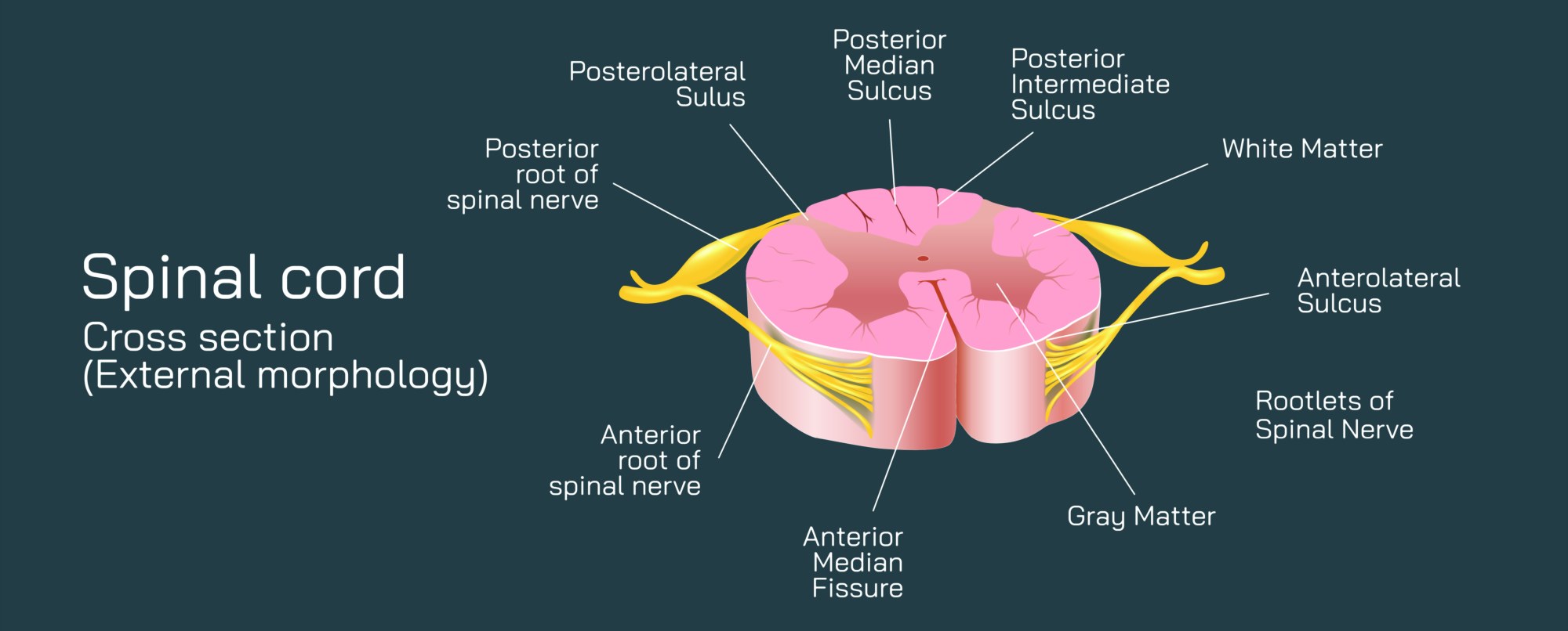
Comprehensive Overview of the Spinal Cord Cross Section and Its External Morphology
The spinal cord is a cylindrical neural structure that extends from the base of the brain through the protective vertebral canal. When viewed in cross section, its external morphology reveals a precise arrangement designed for efficient neural communication and structural support. Understanding this layout helps clarify how sensory and motor pathways are organized and how the cord connects the peripheral system with central processing zones.
Externally, the spinal cord maintains a rounded or slightly oval contour depending on the region. In cervical areas, the outline appears broader due to the high density of ascending and descending fibers. Thoracic regions display a narrower shape, with lumbar segments again widening to accommodate increased nerve supply to the lower limbs. This shifting external form reflects the distribution of neural pathways and structural load at each level.
A defining feature of the external morphology is the presence of longitudinal grooves. The anterior median fissure, a deep indentation on the front surface, separates the cord into two symmetrical halves and contains vessels that supply internal tissue. Opposite this structure lies the posterior median sulcus, a shallower groove on the dorsal side. These landmarks establish clear directional orientation and serve as important reference points for structural organization within the cord.
Surrounding the entire structure is a connective tissue covering made up of protective layers. The dura mater encases the outer region, forming a durable sheath that shields the neural tissue. The arachnoid membrane lies beneath it, providing a delicate barrier, while the pia mater tightly adheres to the cord surface. Between these layers, the subarachnoid space holds cerebrospinal fluid, which cushions the cord and assists with nutrient movement and waste removal.
The spinal cord also displays distinct surface attachments where nerve roots enter or exit. Along the dorsal surface, sensory fibers converge at the dorsal root entry zone. These clusters project into the cord, carrying information from peripheral receptors. On the ventral side, motor fibers exit at the ventral root zone, transmitting signals to muscles and glands. Together, these pathways form segmental units that repeat along the full length of the cord, creating a consistent pattern of sensory intake and motor output.
In addition to these functional zones, the external outline reveals the positions of white and gray matter when the cord is sectioned. While white matter surrounds the exterior, gray matter forms a central butterfly-shaped region. Its orientation can be inferred from the outer contour, as thicker external segments often correspond to increased internal fiber concentration. This relationship between outer shape and internal design highlights how the cord adapts structurally to support complex neural interactions.
Another critical aspect of the external morphology is the presence of the central canal. Though not visible on the outer surface itself, its alignment is defined by the surrounding external shape. It runs longitudinally within the cord and stabilizes fluid movement inside the neural column. Variations in the external outline may subtly shift canal position, especially in regions undergoing changes due to developmental processes or functional demands.
Understanding the spinal cord’s external morphology through cross sectional study offers insight into how neural signals are organized and transmitted. By analyzing grooves, surface landmarks, nerve root zones, and the overall shape at different levels, one gains a clear view of how structure and function work together to maintain communication between the central and peripheral systems. This structural clarity supports clinical diagnosis, anatomical study, and scientific research, forming the foundation for understanding spinal organization and neurological coordination.