Melanin Pigment Colors of Skin Vector Illustration Showing Eumelanin and Pheomelanin Variations Affecting Human Skin Tone Diversity
Melanin is the primary pigment responsible for human skin, hair, and eye color, and it plays a vital role in photoprotection by absorbing ultraviolet (UV) radiation. The two major types of melanin—eumelanin and pheomelanin—determine the wide spectrum of human skin tones and influence susceptibility to sunburn and skin damage. A vector illustration depicting melanin pigment colors typically integrates skin tone variations, pigment types, concentration gradients, and their distribution in the epidermis, providing an educational overview of how melanin contributes to skin diversity. By combining anatomical depiction, color coding, and labeled molecular information, such diagrams make the abstract concept of pigmentation visually intuitive and scientifically accurate.
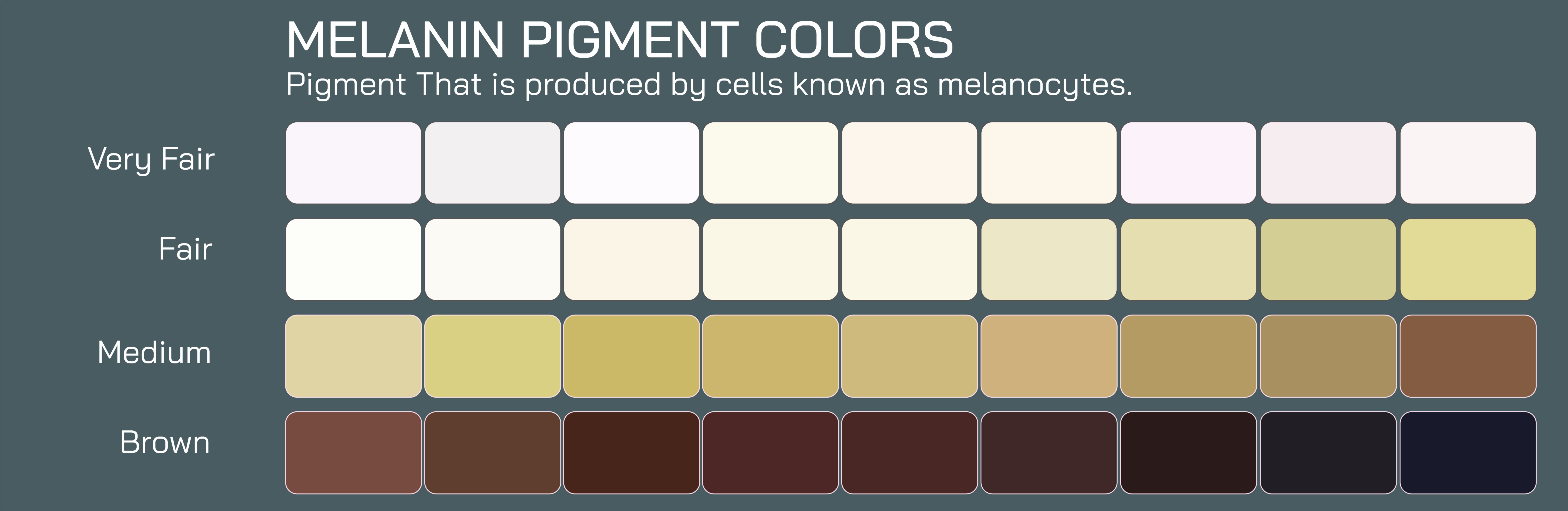
At the core of the illustration is a gradient of human skin tones, ranging from very light to very dark. Each skin tone is labeled to show the predominant type and concentration of melanin present. Eumelanin, responsible for brown and black pigments, is shown increasing in concentration toward darker skin tones, providing UV protection and contributing to deeper hues. Pheomelanin, responsible for red and yellow pigments, is depicted in lighter skin tones, often combined with lower concentrations of eumelanin, producing fair or reddish skin. Color coding or overlay shading visually distinguishes between these two pigments, demonstrating how their relative abundance creates the spectrum of human skin colors.
The vector illustration typically includes a cross-sectional view of the skin, showing the epidermis, dermis, and subcutaneous layers, with melanin concentrated in the basal layer of the epidermis within melanocytes. Arrows indicate the production of melanin within melanosomes and the transfer of pigment to surrounding keratinocytes, highlighting the cellular mechanisms that determine skin color. Labels identify melanocytes, keratinocytes, melanosomes, and pigment types, linking molecular biology to observable skin tone.
Eumelanin distribution is often highlighted in darker skin tones. Vector diagrams show high-density eumelanin granules in the epidermis, illustrating its role in absorbing UV radiation and protecting DNA from photodamage. Arrows may indicate light absorption pathways, emphasizing photoprotective function. Labels such as “high eumelanin concentration → darker skin, enhanced UV protection” provide context for both biological function and observable phenotype.
Pheomelanin is illustrated in lighter or reddish skin tones, often concentrated alongside smaller amounts of eumelanin. Vector diagrams may depict the lighter coloration of pheomelanin granules and their influence on overall skin tone. Arrows or shading may indicate increased susceptibility to UV damage due to lower eumelanin levels, emphasizing the functional differences between pigment types. Labels can highlight characteristics such as “pheomelanin → red/yellow hue, lower UV protection,” connecting pigment chemistry with physiological and evolutionary significance.
A critical feature of the vector illustration is the interaction between eumelanin and pheomelanin in producing intermediate skin tones. Panels may show how variations in pigment ratio and concentration result in olive, tan, and medium brown shades, demonstrating the continuum of human pigmentation. Gradient color bars or pie charts may visually represent the percentage of each pigment type within different skin samples, reinforcing the concept of quantitative variation.
Vector illustrations often include hair and eye color correlations, showing that higher eumelanin produces darker hair and eyes, whereas higher pheomelanin results in lighter or reddish tones. Arrows may connect epidermal melanin to hair follicles and iris pigmentation, demonstrating systemic effects of pigment types. Labels identify melanocyte activity across tissues, highlighting the genetic and biochemical basis for color diversity.
The diagram may also depict adaptive and protective roles of melanin. Darker eumelanin-rich skin is associated with reduced sunburn risk, lower UV-induced DNA damage, and higher vitamin D regulation efficiency in equatorial regions. Lighter pheomelanin-rich skin facilitates vitamin D synthesis in regions with lower UV exposure but increases susceptibility to sunburn and photoaging. Vector arrows and callouts may illustrate UV radiation interactions, linking skin color with environmental adaptation and evolutionary biology.
Additional features in vector diagrams include magnified melanocyte cells showing melanosomes with varying pigment types, arrows illustrating melanin transfer to keratinocytes, and labels highlighting enzymatic control, such as tyrosinase activity, which regulates melanin synthesis. This combination of cellular, tissue, and systemic perspectives provides a complete picture of pigmentation biology.
By integrating skin tone gradients, eumelanin and pheomelanin concentrations, epidermal structure, pigment transfer, and functional significance, a vector illustration of melanin pigment colors provides a holistic understanding of human skin diversity. Color-coded panels, labeled arrows, and cross-sectional diagrams clarify how the relative abundance and distribution of melanin types produce the wide range of human skin tones while connecting these features to UV protection, health, and evolutionary adaptation.
Ultimately, a melanin pigment vector illustration demonstrates the interplay between pigment biochemistry, skin color variation, and physiological function, transforming abstract molecular concepts into a visually accessible and educational tool. Through labeled epidermal layers, pigment granules, concentration gradients, and UV interaction pathways, the diagram allows learners to understand how eumelanin and pheomelanin jointly influence human appearance, photoprotection, and evolutionary adaptation across populations.