Malaria Transmission Cycle Vector Illustration Showing Mosquito Bite Infection Stages and Parasite Life Cycle in Humans
Malaria is a life-threatening disease caused by Plasmodium parasites, which are transmitted to humans through the bite of infected female Anopheles mosquitoes. Understanding the malaria transmission cycle is critical for public health education, disease prevention, and clinical awareness. A vector illustration of the malaria cycle typically integrates mosquito anatomy, human infection stages, parasite development, and transmission pathways, providing a comprehensive visual explanation of how the disease spreads and progresses. By combining labeled vectors, arrows indicating parasite movement, and stage-specific diagrams, such illustrations make complex biological processes accessible and educational.
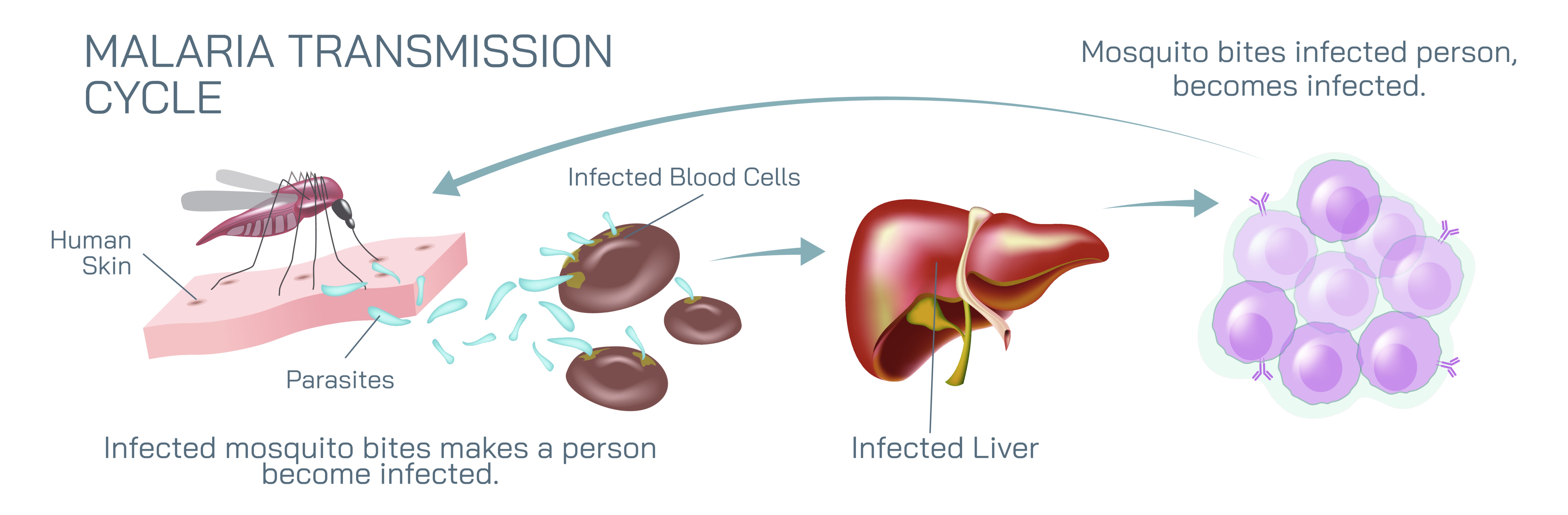
At the center of the illustration is the mosquito vector, commonly depicted as a female Anopheles mosquito in a lateral or dorsal view. Its anatomy is labeled to show key components involved in transmission, such as the proboscis, through which the parasite is injected into humans during a blood meal. Arrows indicate the direction of parasite transfer from the mosquito to the human host, emphasizing the role of the vector in initiating infection. Color coding may highlight infectious stages of the parasite present in the mosquito, such as sporozoites residing in the salivary glands, to demonstrate the readiness of the mosquito to transmit malaria.
The human component of the illustration shows the skin entry point where sporozoites are injected during the mosquito bite. From this entry, arrows lead to the liver, where sporozoites invade hepatocytes and undergo asexual reproduction, forming schizonts. This stage is labeled in the diagram, and magnified insets may depict intracellular parasite growth within liver cells, emphasizing that this pre-erythrocytic stage is asymptomatic but crucial for parasite multiplication. The illustration may also indicate the duration of the liver stage, showing the time delay before parasites enter the bloodstream.
Following liver development, arrows indicate the release of merozoites into the bloodstream, marking the erythrocytic stage, where the parasite invades red blood cells. Vector diagrams often depict red blood cells being infected, with schizonts forming within and subsequently rupturing the cells to release more merozoites. This cyclic destruction of erythrocytes is linked visually to clinical symptoms such as fever, chills, and anemia, providing a connection between microscopic parasite activity and human disease manifestations. Labels may identify parasite forms in red blood cells, including trophozoites, schizonts, and gametocytes, highlighting the stages involved in replication and transmission readiness.
A crucial part of the cycle shown in the vector illustration is the formation of gametocytes, the sexual forms of the parasite, which are taken up by a mosquito during a subsequent blood meal. Arrows indicate the movement of gametocytes from human blood to the mosquito midgut. In the mosquito, gametocytes mature into male and female gametes, undergo fertilization to form zygotes, and develop into ookinetes, which penetrate the midgut wall and form oocysts. These oocysts eventually release sporozoites, which migrate to the mosquito salivary glands, completing the transmission cycle. Color-coded arrows and labeled stages make it clear how the parasite alternates between human and mosquito hosts.
Vector diagrams frequently incorporate side panels or magnified insets for critical stages, such as liver schizonts, blood-stage trophozoites, and mosquito oocysts. These panels can show parasite morphology, intracellular locations, and replication patterns, enhancing understanding of the complex life cycle. Arrows and numbering guide the viewer sequentially through the stages, emphasizing both the cyclical nature of transmission and the dependence on both human and mosquito hosts.
Additional elements often depicted in the illustration include environmental context, such as standing water habitats for mosquito breeding, and preventive interventions like bed nets, insect repellents, and antimalarial drugs. Arrows and labels can indicate how prevention disrupts the transmission cycle, helping learners connect biological processes with public health measures. Highlighting critical stages susceptible to intervention, such as mosquito bites, liver-stage parasites, or gametocyte formation, visually reinforces points of disease control.
By combining mosquito anatomy, parasite stages, human infection sites, blood cell infection, liver stages, gametocyte formation, and vector reentry, a malaria transmission cycle vector illustration provides a thorough and intuitive visualization of the disease process. Color-coded arrows, labeled stages, and magnified insets clarify complex biological interactions and allow viewers to follow the parasite’s journey from mosquito to human and back. The diagram simultaneously illustrates microscopic mechanisms and macroscopic disease transmission, integrating biological, clinical, and ecological perspectives.
Ultimately, a vector illustration of the malaria transmission cycle demonstrates the interconnected roles of human hosts, mosquito vectors, and Plasmodium parasites, providing a visual framework for understanding disease propagation, clinical symptoms, and prevention strategies. Through labeled organs, life stages, directional arrows, and sequential flow, the diagram transforms an intricate parasitic life cycle into an accessible and educational tool, supporting learning for students, healthcare professionals, and public health initiatives.