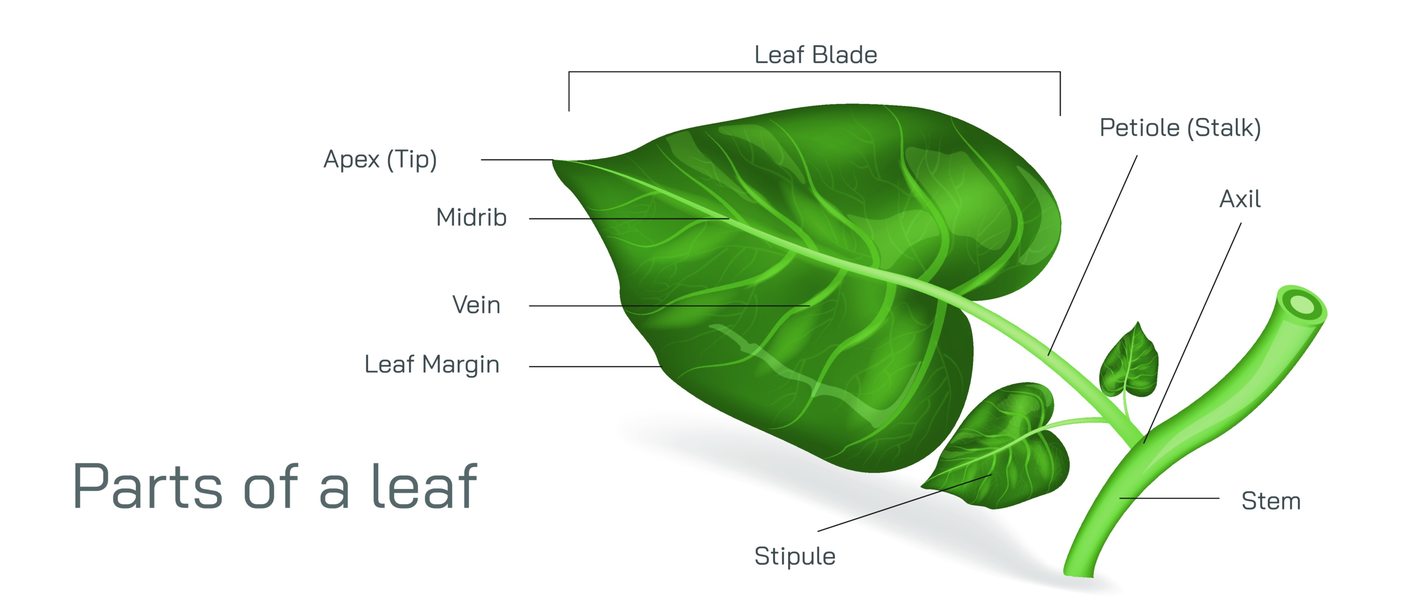
Leaf Anatomy Vector Illustration Showing Internal and External Parts of a Leaf Including Veins, Lamina, Midrib, and Stomata
Leaves are the primary photosynthetic organs of plants, responsible for capturing sunlight, facilitating gas exchange, and supporting water and nutrient transport. Understanding leaf anatomy is essential for botany, plant physiology, and agricultural education. A vector illustration of leaf anatomy typically integrates both external and internal structures, clearly labeling the lamina, midrib, veins, stomata, and supporting tissues, providing a comprehensive and visually intuitive depiction. By combining cross-sectional views, surface structures, vein networks, and microscopic stomatal openings, such illustrations allow learners to understand how structure supports function in leaves.
At the center of the illustration is a full leaf in lateral or top-down view, showing the lamina (leaf blade) as the broad, flat surface. Color coding or shading is used to distinguish leaf tissues and to emphasize regions of interest. Labels highlight the lamina’s role in photosynthesis and light capture, while arrows or overlays indicate its planar area, showing how surface geometry maximizes sunlight absorption.
The midrib, the central thickened vein running longitudinally through the lamina, is illustrated as a prominent structure supporting the leaf. Labels identify it as the primary vascular bundle, containing xylem and phloem, which transport water, minerals, and nutrients. Arrows may indicate the direction of water flow upward through xylem and nutrient transport through phloem, visually linking vascular structure to functional transport. Magnified insets may show the internal arrangement of vascular tissues within the midrib, emphasizing the differentiation between xylem, phloem, and supporting sclerenchyma fibers.
Veins branching from the midrib are illustrated in a hierarchical network, with primary, secondary, and tertiary veins depicted. Labels indicate venation types, such as pinnate or palmate, and show how veins support the leaf structurally while distributing water and nutrients efficiently. Arrows may highlight the direction of sap flow or the pathways of mechanical support, emphasizing the dual functional role of the vascular network. Color coding differentiates veins from surrounding mesophyll, making the network visually distinct and easier to comprehend.
A cross-sectional view of the leaf is included to illustrate internal anatomy. Layers are labeled, including the upper epidermis, palisade mesophyll, spongy mesophyll, and lower epidermis. The palisade mesophyll is depicted with elongated, tightly packed cells rich in chloroplasts for photosynthesis, while the spongy mesophyll shows loosely arranged cells facilitating gas exchange. Labels indicate air spaces within the spongy layer and their role in CO₂ diffusion. Directional arrows may show the movement of gases (CO₂ entering, O₂ exiting) through the leaf, highlighting functional integration.
Stomata are depicted on the lower epidermis, shown as microscopic openings flanked by guard cells. Labels indicate their role in gas exchange and transpiration, with arrows showing the entry of carbon dioxide and the exit of oxygen and water vapor. Magnified insets illustrate the stomatal aperture and guard cell mechanics, connecting microscopic structures to macroscopic leaf function. Color coding may distinguish open versus closed stomata, visually reinforcing their regulatory role in response to environmental conditions.
Vector diagrams may include leaf hairs (trichomes), cuticle, and wax layers, highlighting external protective features. Arrows indicate water retention and UV protection functions, linking structural adaptations to environmental resilience. Insets may show cuticle thickness or trichome types, enhancing educational value for detailed morphological study.
Additional features often include comparative panels showing different leaf types, venation patterns, or anatomical adaptations for varied environmental conditions. Arrows and labels may indicate variations in stomatal density, mesophyll thickness, or vein prominence, providing a broader understanding of leaf functional diversity.
By combining lamina, midrib, veins, epidermis, mesophyll, stomata, and protective structures in a color-coded, labeled vector illustration, learners gain a holistic understanding of leaf anatomy. Arrows, magnified insets, and cross-sectional views clarify both internal and external features, linking anatomical structures to physiological functions like photosynthesis, gas exchange, and water transport.
Ultimately, a vector illustration of leaf anatomy demonstrates the integration of structural components that enable photosynthesis, support, and transport, transforming abstract botanical concepts into a visually engaging, educational, and intuitive tool. Through labeled veins, midrib, lamina, stomata, and tissue layers, the diagram provides a comprehensive reference for botany students, plant physiologists, and educators.