THE COMPLEX STRUCTURE OF THE HEEL AND FOOT: AN IN-DEPTH ANATOMICAL AND FUNCTIONAL OVERVIEW
The heel and foot together form one of the most extraordinary architectural masterpieces of the human body because they serve as both a platform for weight bearing and a dynamic mechanism for movement. They support every shift of posture, every step in the gait cycle, and every physical action that depends on balance or locomotion. Although the foot appears small when compared to the rest of the body, the precision and strength built into its design are remarkable. The heel, located at the rear of the foot, operates as the primary point of initial contact with the ground during walking. It has the critical responsibility of receiving the body’s downward force and transforming it into mediated impact that the rest of the body can tolerate safely. Without this remarkable system of shock absorption, even the simple act of standing upright would place overwhelming stress on the skeletal and muscular frameworks above it.
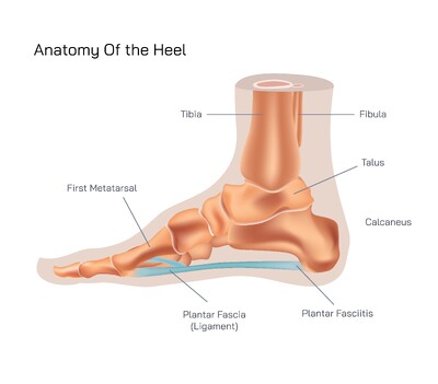
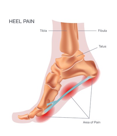
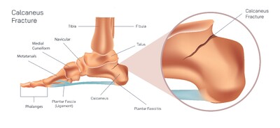
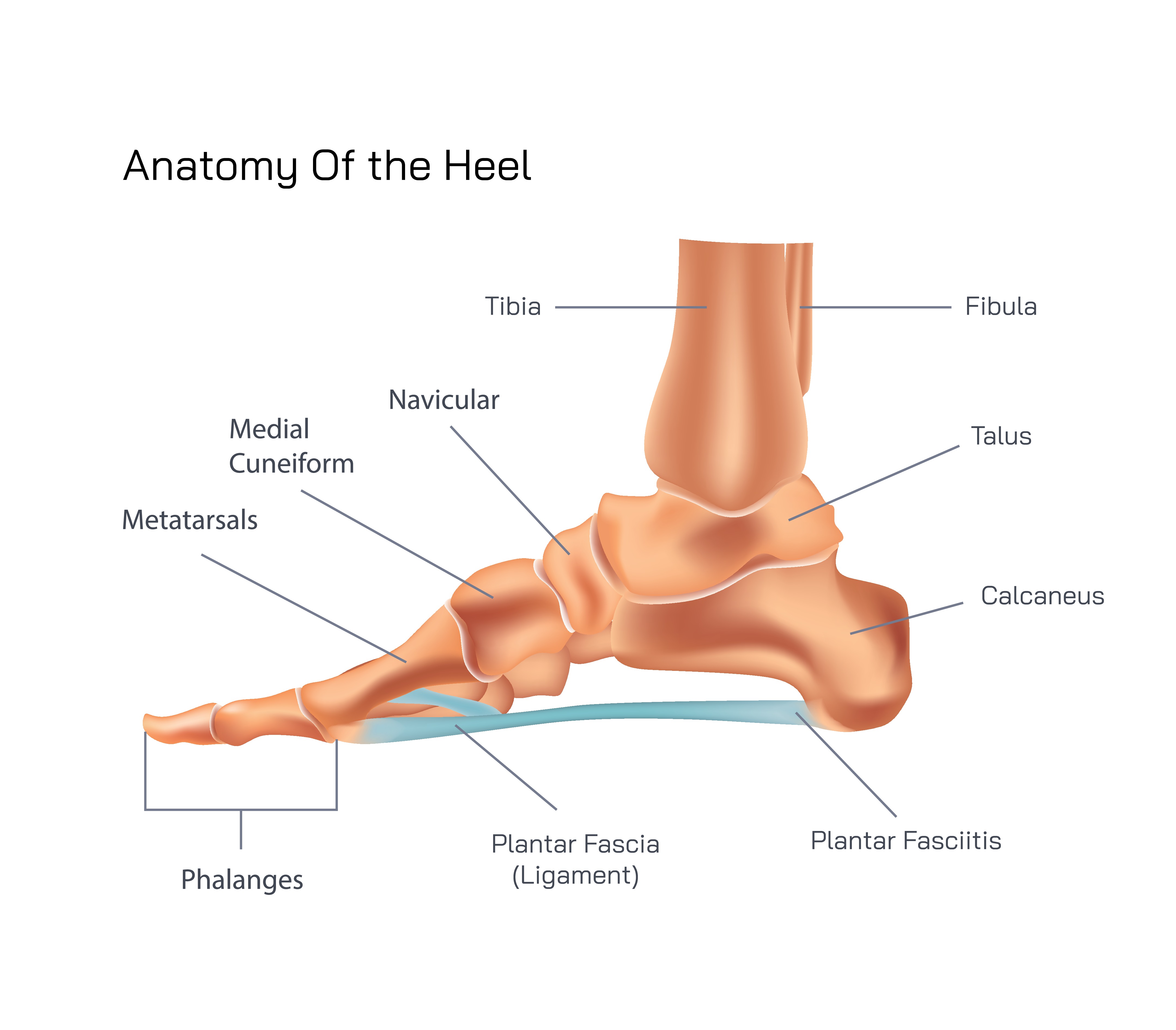
The foundation of the heel is the calcaneus bone, which is uniquely shaped and positioned to bear weight and resist compressive forces. It connects to the talus at the subtalar joint, a specialized junction that enables the foot to adapt to different surfaces by tilting inward or outward when necessary. This anatomical arrangement allows the foot to adjust to uneven terrain, contributing greatly to stability with every step. On the underside of the heel lies the heel fat pad, an incredibly important cushioning structure composed of dense compartments filled with fibrous connective tissue and fat. It acts like a natural shock-absorbing device, compressing on impact and springing back into shape repeatedly. The durability of this system is extraordinary, as it must withstand thousands of impacts every single day.
Forward from the heel, the bones of the foot extend in a precise arrangement that creates the hindfoot, midfoot, and forefoot. These three divisions function together as one coordinated system. The hindfoot consists of the calcaneus and talus and forms the structural base. The midfoot contains the navicular, cuboid, and cuneiform bones, contributing to the arches that give the foot its elasticity and spring. The forefoot includes the metatarsals and phalanges, which form the toes and front of the foot, responsible for push-off during the gait cycle. Instead of being flat, the inner structures of the foot are shaped like a three-dimensional suspension bridge that uses arches to distribute force across multiple areas rather than concentrating it in one location. The medial longitudinal arch is the tallest and most critical for weight distribution, the lateral longitudinal arch stabilizes the outer side of the foot, and the transverse arch stretches across the midfoot to maintain width and balance. These arches act as mechanical energy storage systems, loading and unloading force during movement to create efficiency and minimize fatigue.
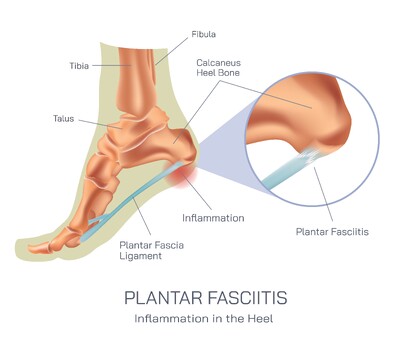
Supporting this bony framework is an intricate network of muscles, tendons, and ligaments. The Achilles tendon is the strongest and thickest tendon in the human body and connects the calf muscles to the calcaneus. Through this connection, the lower leg and foot operate as a single kinetic chain that can raise the heel, allowing propulsion required for walking, climbing, and running. The foot also contains intrinsic muscles located entirely within its structure, which help control the fine movements of the toes and stabilize the arches, while extrinsic muscles originate in the leg and insert into the foot, controlling major movements such as flexion and extension. Ligaments bind the bones tightly together, preventing injuries and ensuring the structural configuration remains intact despite major stress. The plantar fascia, a broad band of connective tissue stretching from the heel to the toes, acts like a bowstring, supporting the medial arch and resisting forces that would flatten the foot.
The heel and foot also depend on their sensory and circulatory systems to function correctly. Nerves provide constant information about pressure, balance, and movement, feeding data to the brain so the body can make micro-adjustments instantly to avoid falling or losing stability. Blood vessels deliver oxygen and nutrients and remove waste, ensuring tissues remain healthy and capable of enduring continuous stress. When issues disrupt this harmony, the symptoms are immediately noticeable because the heel and foot are responsible for foundational tasks that no other part of the body can compensate for. Conditions like plantar fasciitis, Achilles tendonitis, flat feet, high arches, or heel spurs can dramatically affect mobility and overall quality of life.
The structure of the heel and foot represents a stunning combination of stability, flexibility, strength, and fine motor control. Its role in human life is unmatched because it serves as the physical base that carries the full weight of the body while performing complex coordinated motions. Whether one is standing still, sprinting forward, climbing uneven ground, or simply shifting posture, the heel and foot work continuously without conscious effort. Understanding their design is essential not only for anatomical study but also for fields such as podiatry, sports science, orthopedics, rehabilitation, and everyday health. The harmony between bones, muscles, connective tissues, nerves, and vascular structures demonstrates how evolution engineered a support system capable of both resilience and graceful movement.