The anatomical structure of the foot represents one of the most remarkable engineering achievements in the human body. Although small in size compared to the rest of the skeletal system, the foot supports the entire body weight while standing and absorbs tremendous forces during walking, running, jumping and everyday movements. The foot is not a single solid object but a complex network of bones, muscles, tendons, ligaments, connective tissues, arches, joints, nerves and blood vessels that work together seamlessly to maintain balance, provide stability and generate movement. Whether the body is at rest or in motion, the foot plays an essential and continuous role in ensuring mobility and structural support.
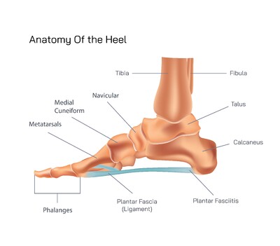
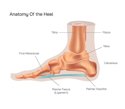
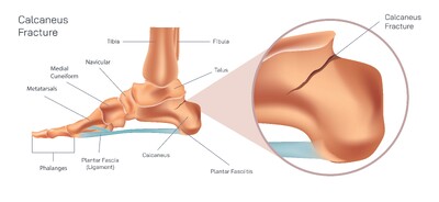
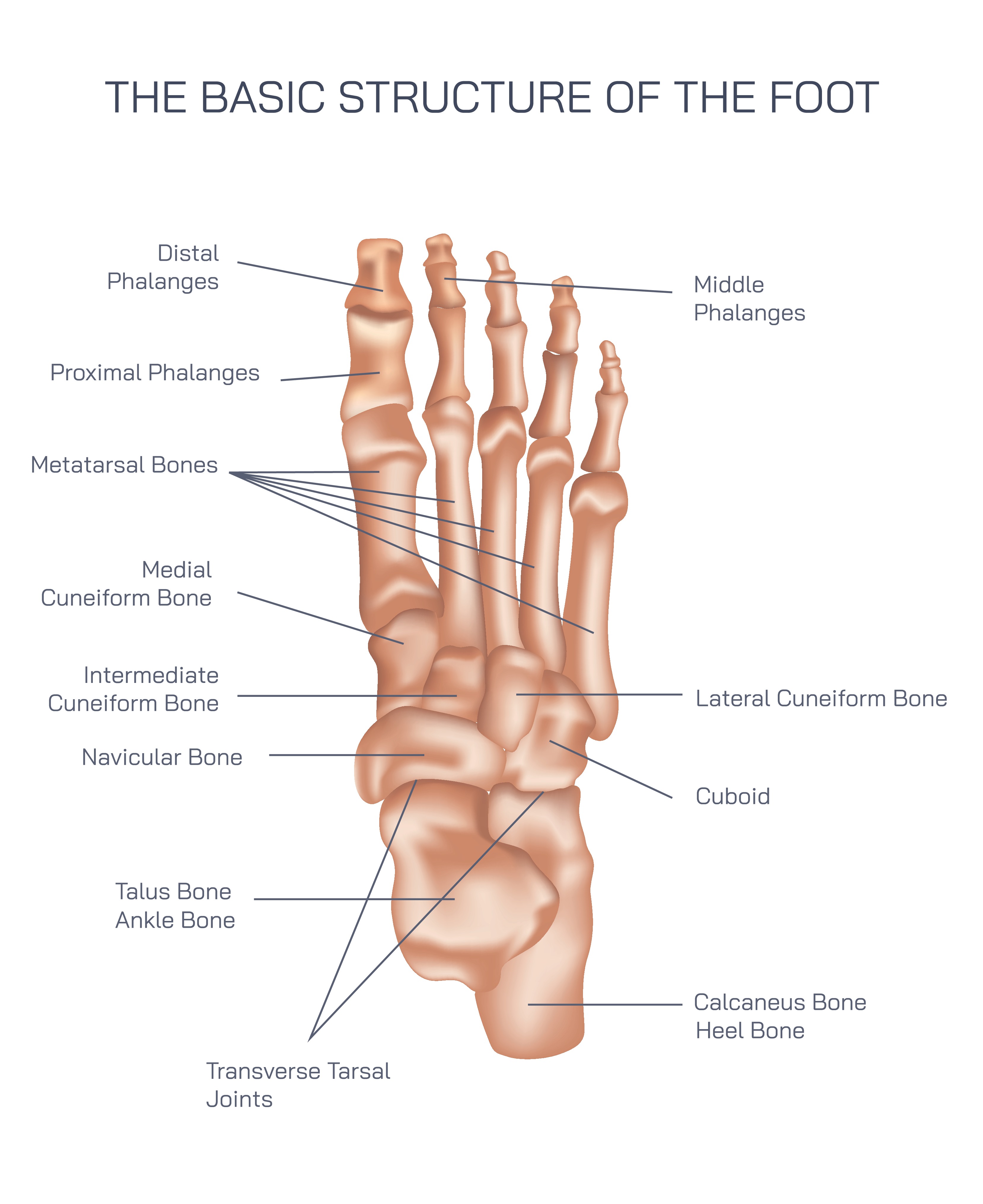
At the core of the anatomical structure of the foot lies its skeletal foundation, consisting of 26 bones organized into three major regions: the hindfoot, midfoot and forefoot. The hindfoot contains the calcaneus (heel bone) and talus. The calcaneus bears much of the body’s weight and acts as the point of initial contact during walking. The talus sits above the calcaneus and forms the lower component of the ankle joint, connecting the foot to the lower leg. These bones establish the core support that withstands the first impact when the foot strikes the ground. The subtalar joint, formed between the talus and calcaneus, enables the foot to tilt inward or outward, helping the body adapt to uneven terrain and maintain balance.
The midfoot is the structural bridge between the hindfoot and forefoot and contains the navicular, cuboid and three cuneiform bones. These bones form the central support of the arches and allow the foot to act as a flexible yet sturdy platform during movement. When the body shifts forward while walking, the midfoot transfers force from the heel to the forefoot, distributing pressure evenly and preventing excessive stress on any single area. The forefoot consists of the five metatarsal bones and the 14 phalanges forming the toes. Although often taken for granted, the toes are essential in generating propulsion; when the heel lifts off the ground, the forefoot pushes the body forward through coordinated extension of the toes.
The arches of the foot are a defining component of its anatomy and are responsible for the foot’s exceptional ability to bear weight and absorb shock. The medial longitudinal arch runs along the inner border of the foot and is the highest and most elastic. It stores mechanical energy and releases it during movement to make walking and running more efficient. The lateral longitudinal arch runs along the outer side of the foot and provides additional stability. The transverse arch spans across the midfoot and helps maintain the foot’s structural shape while adapting to varying surfaces. When functioning correctly, these arches protect the body from repetitive impact and ensure smooth, balanced motion. When weakened, flattened or excessively rigid, they can contribute to foot pain, instability and altered gait.
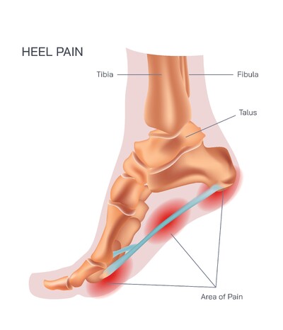
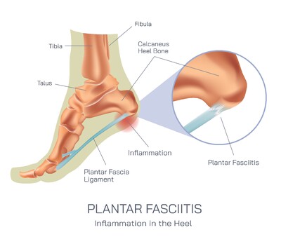
Muscles and tendons play a dynamic role in foot anatomy. The extrinsic muscles originate in the lower leg and extend into the foot, producing gross movements such as pointing the toes, lifting the foot and raising the heel. The intrinsic muscles lie entirely within the foot and provide fine motor control by stabilizing the toes and maintaining the shape of the arches. The Achilles tendon, which connects the calf muscles to the heel bone, is critical for propulsion because it enables the heel to lift during walking and running. The plantar fascia, a thick band of connective tissue running from the heel to the toes, supports the medial arch and reinforces the structural integrity of the foot during every step. When overstressed, the plantar fascia may become inflamed, causing plantar fasciitis — one of the most common sources of heel pain.
Ligaments serve as stabilizers by holding the bones in proper alignment and preventing excessive motion within the joints. These ligaments give the foot both rigidity and controlled flexibility, ensuring that movements remain smooth and safe rather than unstable or erratic. Joints within the foot — including the ankle joint, subtalar joint and numerous intertarsal and metatarsophalangeal joints — allow complex yet coordinated motion. Without these joints working in synergy, basic actions such as walking and balancing would be nearly impossible.
The neurological and circulatory systems complete the anatomical structure of the foot and are essential for functional performance. Nerves distributed throughout the foot provide sensory feedback required for balance and posture. Every shift in ground texture, pressure or slope is immediately detected and communicated to the brain, triggering micro-adjustments that optimize stability. This allows people to walk confidently without constantly watching the ground. Blood vessels supply oxygen and nutrients to the muscles and tissues while supporting recovery from daily wear and tear. A healthy vascular system in the foot is vital for tissue repair and endurance.
While the foot is inherently strong, its anatomical structure remains vulnerable to overload, misalignment and improper support. Excessive standing, high-impact sports, inadequate footwear, sedentary behavior, muscle weakness and aging can all disrupt the natural balance of the foot. These disruptions may lead to common issues such as plantar fasciitis, Achilles tendon irritation, arch collapse, metatarsalgia or stress fractures. The interconnected nature of foot anatomy means that a problem in one region often affects other parts of the body. For example, flat feet may lead to knee misalignment, hip strain or lower back pain over time.
Maintaining the anatomical health of the foot requires awareness and proactive care. Supportive footwear, stretching routines, intrinsic foot muscle strengthening, maintaining healthy body weight and avoiding sudden increases in physical intensity help preserve the natural function of the foot. People who understand the structure of their feet can make informed decisions that reduce injury risk and improve performance in daily life and athletic activity.
The anatomical structure of the foot is a masterpiece of natural biomechanical design. It supports the body during stillness, stabilizes posture, adapts to varied surfaces and powers movement through coordinated muscle action, joint articulation and arch elasticity. When the structures of the foot are nurtured, strengthened and protected, they provide a lifetime of mobility, comfort and physical independence. By appreciating the complexity and importance of the foot’s anatomy, individuals can take steps — in both the literal and figurative sense — toward lasting musculoskeletal health.