3D Neuron Illustration: A Deep Exploration of Its Structure and Functional Importance
A three dimensional rendering of a neuron brings one of the most remarkable biological units to life. The neuron is not only the basic working unit of the brain but also a carefully crafted communication element that maintains the flow of information throughout the nervous system. When viewed in 3D, its shape reveals a fascinating network of branches, extensions, and connections that work together to carry signals to muscle fibers, gland cells, and neighboring nerve cells. This detailed perspective highlights the extraordinary way structure supports purpose.
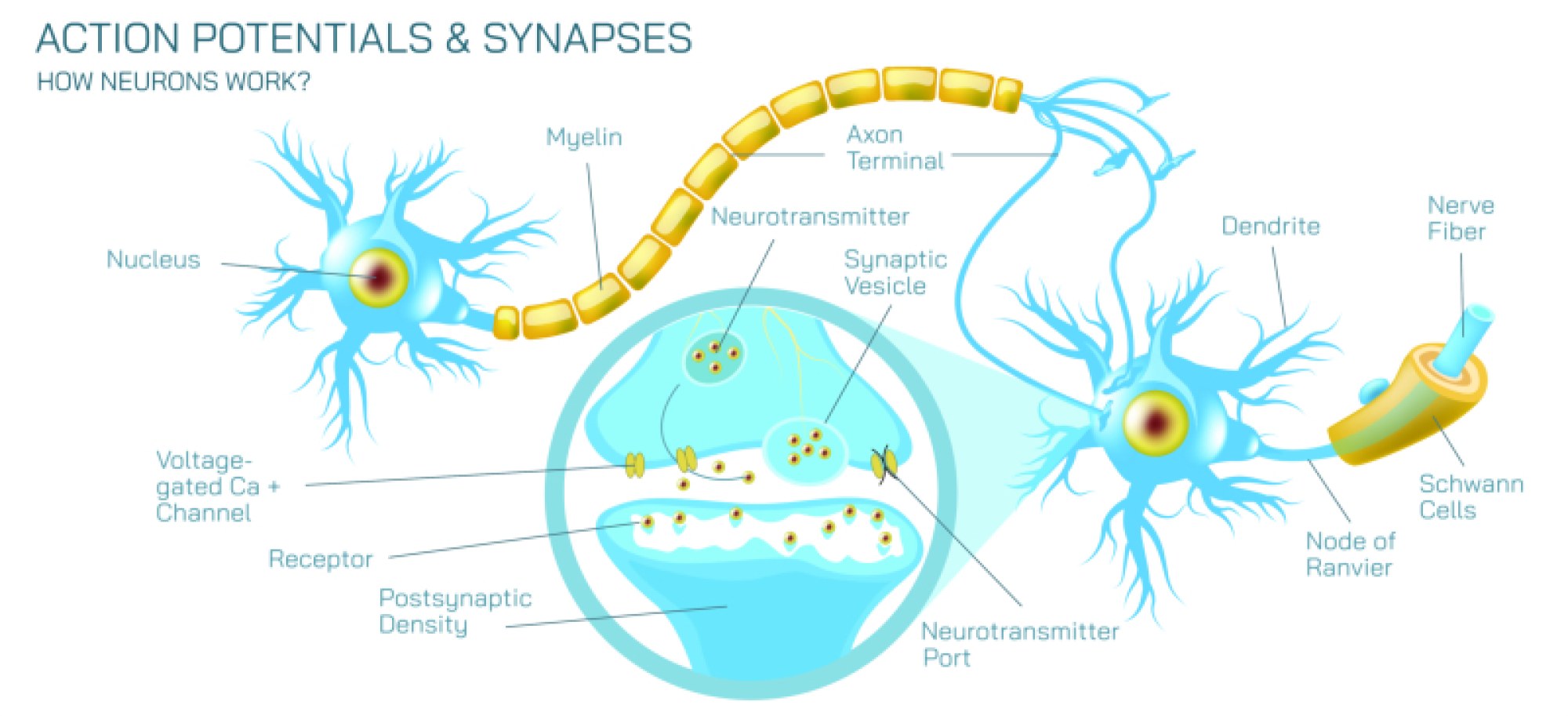
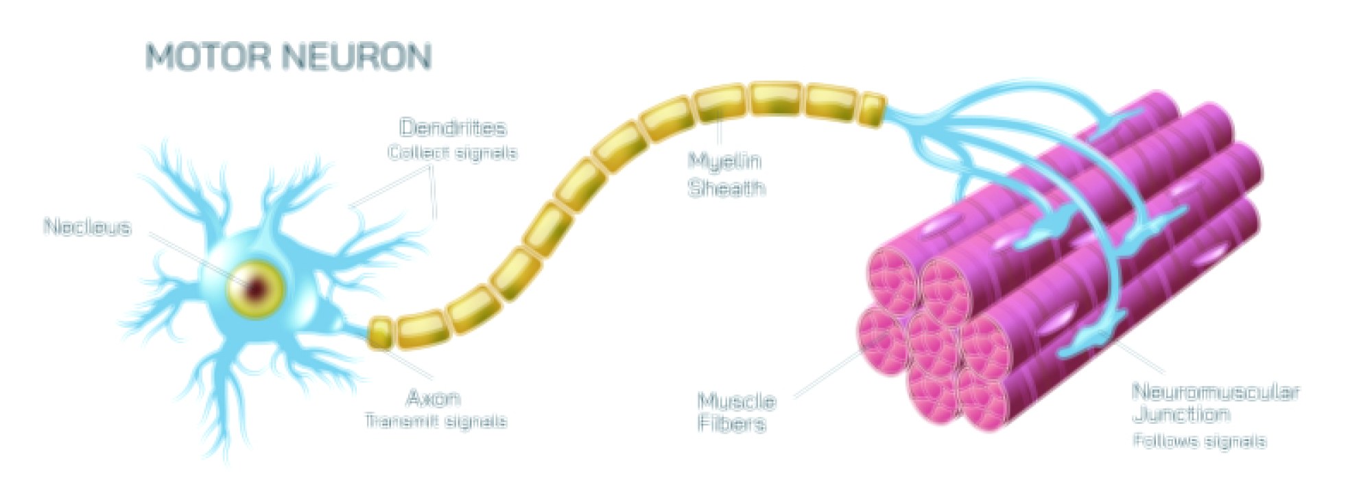
In the 3D model, the most striking feature is the collection of branching extensions called dendrites. These appear like delicate arms reaching outward in multiple directions, forming a wide surface area that invites incoming signals. They resemble fine tree branches, giving the neuron an open shape designed for receiving information. Their irregular spread helps the neuron gather signals from many surrounding cells simultaneously, allowing complex patterns of communication to occur within the nervous system.
At the center of the illustration sits the soma, or cell body, which appears as a rounded hub. It contains the essential machinery that keeps the neuron alive and functioning. The 3D rendering reveals a smooth outer surface and a slightly glowing internal core, suggesting its role as a processing center. All incoming signals enter this region before the cell decides whether to carry the information forward. This central location makes the soma a vital intersection that integrates numerous neural messages.
Extending from the soma is the axon, a long, slender projection that often appears as a smooth tubular line stretching across the model. The 3D view highlights its length and elegance, showing how it acts as the neuron’s primary output path. In many illustrations, segments along the axon appear slightly bulged or separated, representing supportive coverings that help maintain signal strength. The length of the axon demonstrates how a neuron can send information across significant distances, linking the brain to muscles, organs, or distant nerve networks.
The axon terminates in a set of branching endings that resemble a spreading fan. These endings form connection points where signals are passed to other cells. In the 3D rendering, these regions appear as glowing bulb-like structures, symbolizing communication zones where incoming electrical activity transforms into chemical messages. These endings allow the neuron to influence multiple targets, creating widespread effects from a single processed signal.
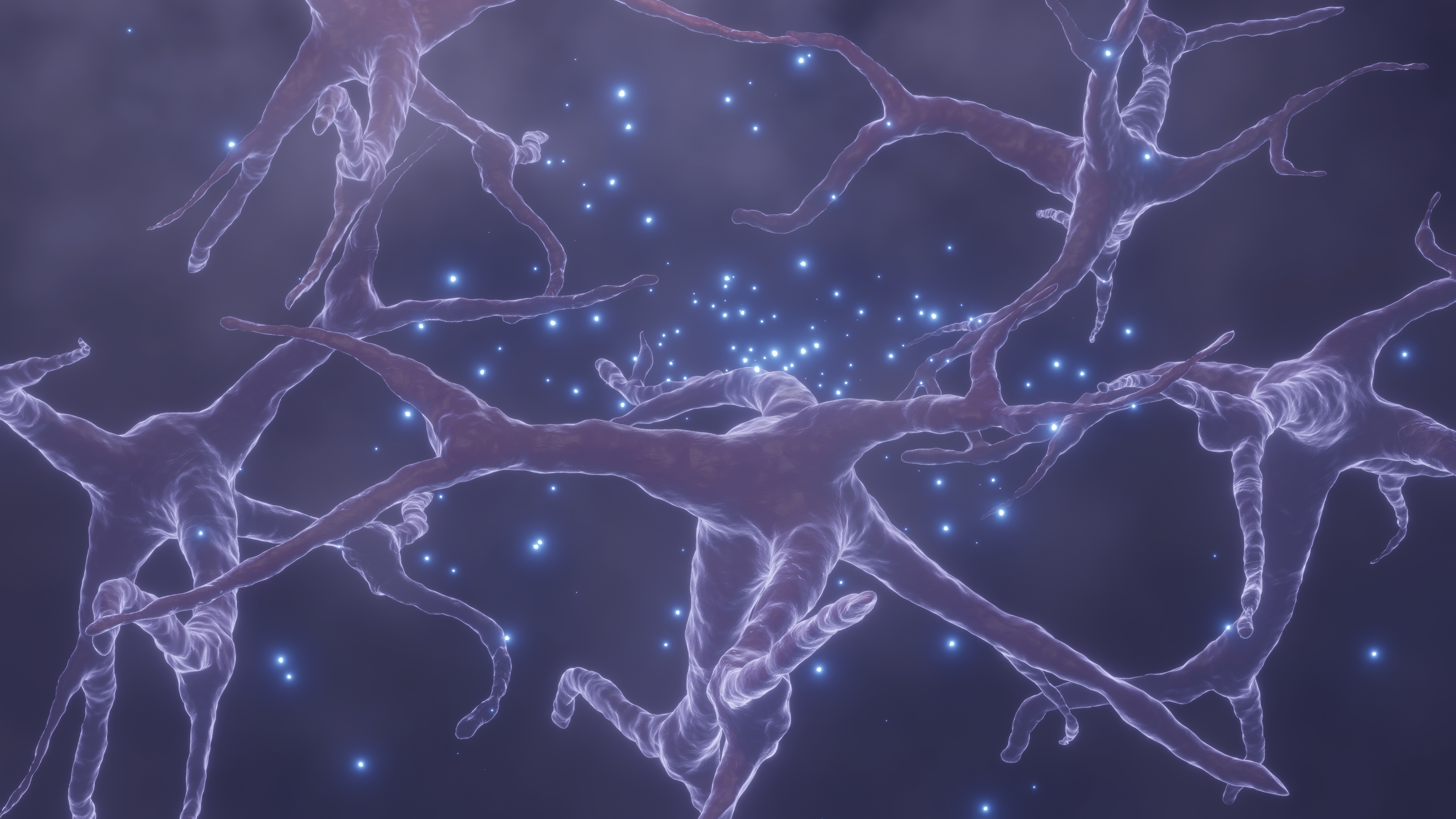
The synapse, while microscopic, becomes visually meaningful in a 3D model. It is portrayed as a small gap where two cells face each other without touching. Bright points or subtle light flows often represent the moment when the signal crosses this gap. This visual cue helps learners understand that communication in the nervous system is not always a direct physical link but a coordinated exchange across tiny spaces.
Another important feature often emphasized in 3D graphics is the interconnectedness of neural pathways. Neurons rarely operate alone. Instead, they form large networks where signals travel in multiple directions. The illustration highlights this by showing numerous dendrites receiving messages and multiple axon endings sending them. This dynamic arrangement reflects how thoughts, actions, memory, and sensory experiences arise from countless coordinated exchanges supported by these cells.
The 3D perspective also helps reveal the neuron’s role beyond basic communication. By examining its structure from different angles, viewers can appreciate how the cell adapts to its environment. Long axons allow distant interaction, while short, dense dendrites enhance local communication. These variations show how neurons specialize depending on their location and function, whether they guide movement, sense external stimuli, or coordinate internal processes.
A fully rendered neuron also emphasizes the elegance of biological engineering. Every shape, branching pattern, and surface detail has meaning. The more viewers explore, the more they understand how this cell allows the brain to think, respond, interpret, and adapt. Its form is both artistic and purposeful, revealing the deep relationship between structure and experience.
Ultimately, a 3D rendered neuron is more than an educational image. It is a window into the complexity of human function. By observing it in detailed form, learners gain a richer appreciation for the invisible communication system that supports movement, thought, emotional expression, and vital responses. The neuron stands as a shining example of how tiny biological elements create the vast landscape of human behavior and perception.