Understanding Microscopic Bacterias and Infections Through 3D Rendered Visualization
A 3D rendered illustration of microscopic bacterias and infections provides an engaging, detailed view of biological processes that are otherwise invisible to the naked eye. These tiny organisms, though small in size, play a major role in human health, either maintaining balance in natural systems or causing disease when uncontrolled. By presenting bacterias in a volumetric 3D environment, viewers gain a better understanding of their structure, behavior, and interactions within tissues or laboratory cultures, making complex scientific concepts easier to grasp.
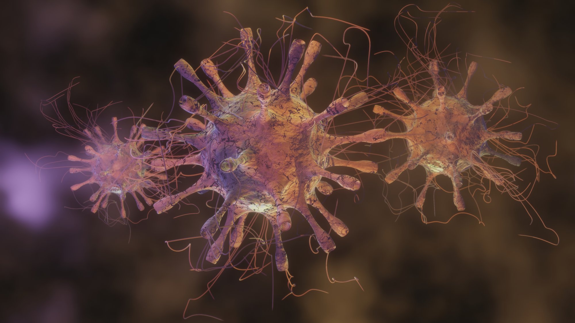
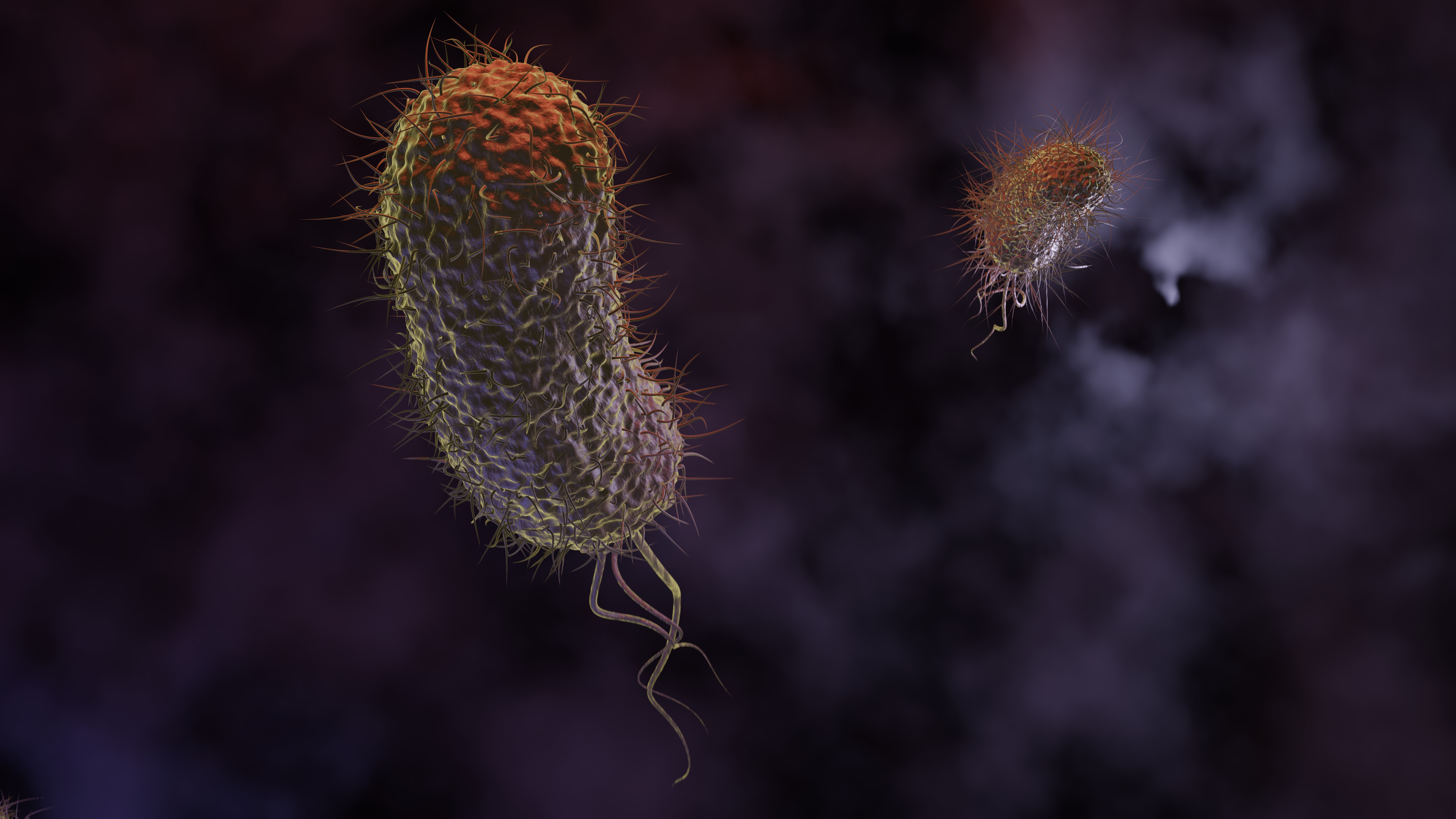
The illustration typically highlights bacterial shapes—spherical (cocci), rod-shaped (bacilli), or spiral (spirilla)—alongside textured surfaces and appendages like flagella or pili. This attention to microscopic detail helps learners visualize how bacterias attach to host cells, multiply, and form colonies. The addition of surrounding environments, such as fluid pathways or cellular matrices, provides context to their movements, showing how infections spread and interact with their hosts.
Infections often begin with bacterial colonization in a vulnerable tissue or environment. A 3D rendered scene can show initial attachment points, clusters forming, and gradual population growth. These visuals demonstrate why early-stage infections can go unnoticed and why prompt medical intervention is essential. Realistic lighting and shadows accentuate contours, textures, and spatial relationships, enhancing comprehension of how pathogens occupy and navigate the microscopic world.
Furthermore, the illustration can depict interactions between bacterias and immune defenses. Elements like white blood cells or molecular markers can be included to show the body’s natural response. This adds an educational layer, emphasizing the battle between pathogenic organisms and host defenses. By visualizing both sides of the process, students and professionals can better understand infection mechanisms, immune strategies, and potential outcomes.
The 3D rendering also emphasizes colony formation and the structural arrangement of bacterial communities. Bacteria rarely exist as isolated cells; they organize into microcolonies, biofilms, or clusters. These formations are critical to their survival and virulence, and showing them in 3D makes it easier to grasp their functional significance. The visual representation helps illustrate how bacterial density, spatial organization, and surface interaction contribute to infection persistence.
Educationally, such a detailed 3D illustration bridges the gap between abstract microbiology concepts and tangible learning. Students, researchers, and healthcare professionals benefit from seeing pathogens in a realistic, dimensional space rather than flat diagrams. It enhances memory retention, understanding of microbial behavior, and comprehension of disease progression. Additionally, the visual is valuable for presentations, textbooks, research publications, and public health awareness materials, translating complex scientific ideas into clear, informative visuals.
The aesthetic elements of a 3D render—lighting, color gradients, and surface textures—enhance engagement, making microscopic bacterias appear both scientifically accurate and visually compelling. This attention to realism ensures that the illustration is not only educational but also immersive, allowing viewers to explore the tiny world of microorganisms as if under a virtual microscope.
Overall, a 3D rendered microscopic bacterias and infections illustration provides a powerful learning tool. It demonstrates the form, function, and behavior of bacteria in relation to infections, immune responses, and environmental interactions. By combining scientific accuracy with visual clarity, it transforms unseen microscopic processes into a comprehensive, engaging, and educational experience for audiences ranging from students to medical professionals.