3D Rendered Virus Illustration Showing Infection and Cellular Hijacking
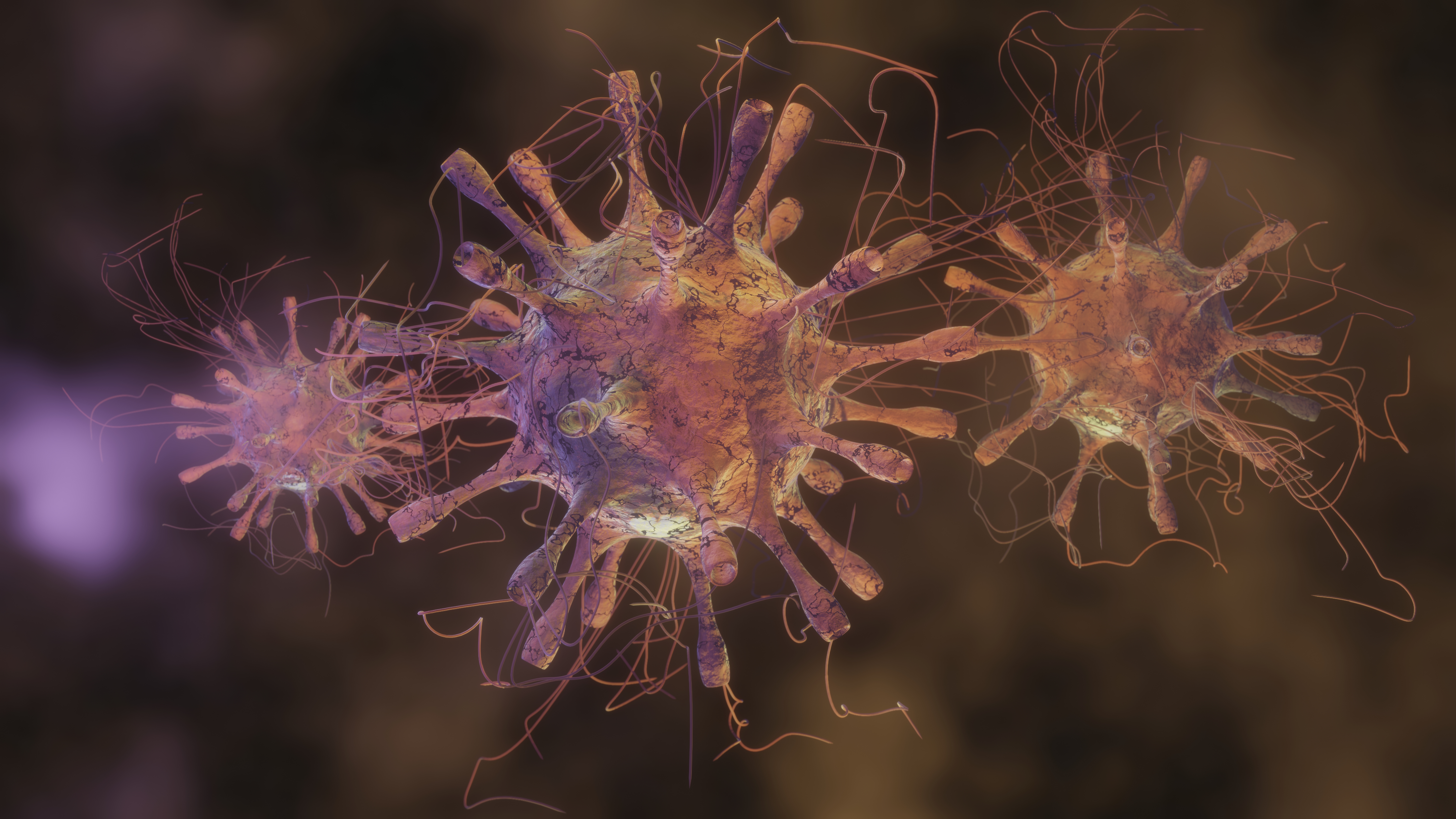
A 3D rendered illustration that represents how viruses infect a host cell brings a deeply complex biological process into a visually accessible format. Understanding viral infection requires an appreciation for the sophisticated interaction between a virus and the cell it targets. A detailed 3D representation reveals each stage clearly, allowing viewers to visualize how a virus attaches to the cell surface, penetrates the membrane, releases its genetic material and begins the takeover of the cell’s internal machinery. This transformation from an inactive particle into a fully active biological invader forms the core of the viral life cycle, and a refined 3D artwork conveys that transition with clarity.
The infection process begins when a virus encounters a compatible host cell. The outer structure of many viruses contains surface proteins that enable them to attach only to specific receptors found on certain types of cells. A 3D illustration highlights these surface proteins, often depicted as spikes or complex molecular shapes extending from the viral shell. This detailed rendering helps viewers understand how each spike binds to a matching receptor on the cell membrane, creating a lock-and-key interaction that initiates entry. This is a critical step, as the virus cannot proceed without successfully attaching to a suitable host.
Once attachment is complete, the virus begins to breach the cell’s protective barrier. A high-quality 3D illustration can show how the viral envelope fuses with the cell membrane, or how the virus injects its genetic material directly into the cell interior. This moment marks a dramatic shift, as the viral genome enters the cytoplasm and seeks out the cell’s existing molecular tools. The contrast between the structured viral particle and the fluid interior of the cell becomes visually striking, emphasizing the transition from an external organism to an internal infiltrator.
Inside the cell, the viral genetic material begins to exert its influence. A 3D depiction can show how the viral genome interacts with ribosomes, enzymes and other cellular components. The cell, originally dedicated to maintaining its own health, now becomes an unintended factory for producing viral proteins and assembling new particles. A rendered visualization often highlights the reorganization of internal structures, presenting the cellular environment as increasingly dominated by viral components. The shift from normal cellular activity to viral manufacturing becomes a central theme of the artwork, showing the gradual takeover with precision and depth.
As replication progresses, the newly formed viral proteins and genomes come together to create complete virus particles. A 3D visualization can capture this assembly process, revealing how individual components converge to form fully structured viral bodies. Within the cell, clusters of new viruses begin to accumulate, creating a sense of increasing pressure. This visual buildup communicates the destructive nature of viral replication, as the cell becomes overwhelmed by the very invaders it unintentionally produced.
When the replication cycle reaches its peak, the cell reaches a breaking point. The newly assembled viruses may exit the cell through controlled release or burst out in large numbers. A 3D illustration often uses dynamic lighting, particle effects and structural fragmentation to show how these viruses emerge, ready to infect neighboring cells. This final release stage completes the narrative of hijacking and destruction, demonstrating how a single viral particle can give rise to countless progeny, each capable of repeating the cycle.
The value of a 3D rendered depiction lies in its ability to blend scientific accuracy with visual storytelling. It highlights the elegance of viral structure, the vulnerability of living cells and the dramatic transformation that unfolds during infection. Through smooth shading, depth effects, dynamic color gradients and meticulous detailing, the illustration conveys both the complexity and the urgency of viral behavior. Whether used in medical education, scientific presentations, health awareness campaigns or research materials, such a graphic becomes an essential tool for understanding a microscopic conflict that shapes many aspects of human health.
A detailed 3D rendering therefore transforms the abstract concept of viral infection into a vivid, identifiable sequence. By visually mapping attachment, entry, genetic release, cellular hijacking, replication and viral emergence, it provides a comprehensive view of how viruses use the host cell as a tool for their own survival. This form of visual communication deepens understanding and fosters clarity in exploring the dynamic world of microscopic pathogens.