Understanding Microscopic Bacterias and Infections Through a 3D Rendered Illustration
A 3D rendered illustration of microscopic bacterias and infection stages provides an exceptionally clear way to visualize biological processes that are otherwise invisible to the naked eye. This type of visual helps learners, educators, medical professionals, and researchers understand how tiny organisms initiate and spread infections within living systems. With depth, texture, and realistic lighting, the render allows viewers to see structural details such as cell walls, surface proteins, and surrounding environments, offering a more complete understanding of microbial behavior.
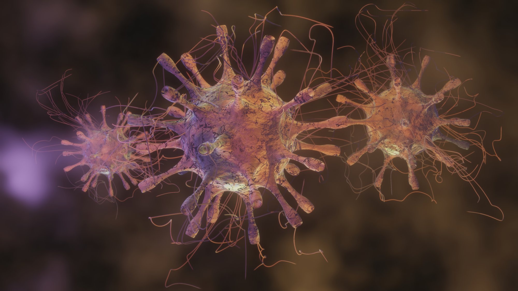
Microscopic bacterias operate at scales far smaller than human perception, making them ideal subjects for 3D representation. The illustration often shows rounded, rod-shaped, or spiral structures that represent different bacterial forms. These shapes are enhanced with realistic textures to highlight surface layers or protective coatings. This visual accuracy helps viewers understand how these organisms attach to surfaces, invade tissues, or replicate inside hosts. The 3D environment may also include floating particles that represent toxins, molecular debris, or immune responses, showing how infections progress in dynamic systems.
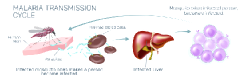
The depiction of bacterias in a 3D scene also helps explain the infection cycle. Typically, an infection begins when bacterias come into contact with a host surface. The illustration might show the organisms clustering around a vulnerable point, emphasizing the initial stages of colonization. By presenting this process with realistic lighting and motion cues, the 3D visual communicates how rapidly bacterias can multiply once they find a suitable environment. This type of clarity is particularly important in teaching how infections grow and why early prevention plays a vital role in healthcare.
As the infection spreads, the render can highlight interactions between bacterias and the immune system. The visual may show white blood cells attempting to neutralize invading organisms, demonstrating the body's natural defense mechanisms. This helps viewers grasp the balance between microbial aggression and immune resistance. It also reinforces the concept that infections occur when bacterias overwhelm these defenses or produce harmful substances that disrupt normal cellular activity.
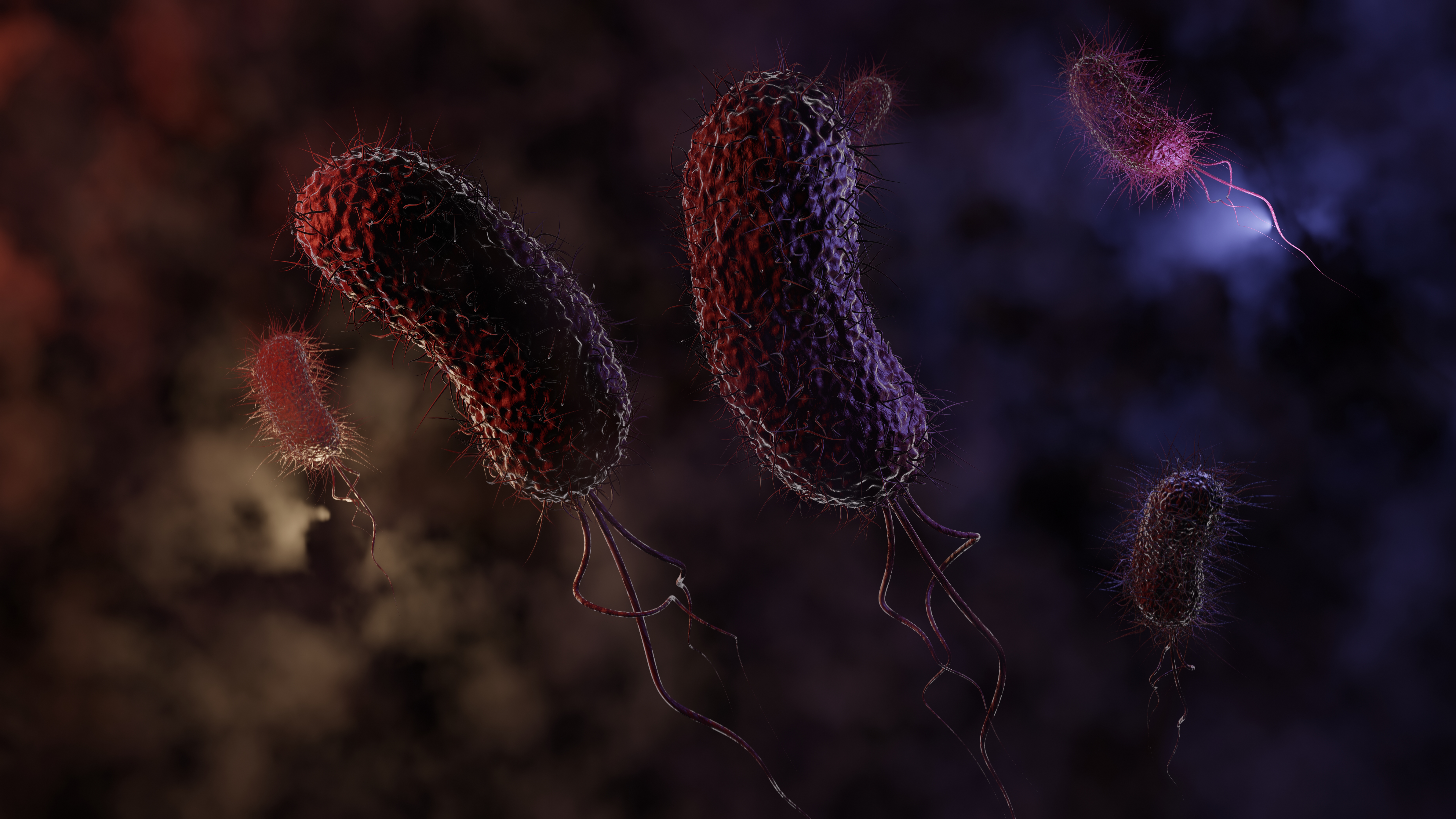
A 3D depiction also illustrates the structural complexity of bacterias. Many organisms possess specialized appendages such as pili or flagella, which help them move or attach to surfaces. In a rendered environment, these structures become highly visible, making it easier to understand how bacterias adapt to different conditions. This clarity is especially valuable for educational and diagnostic purposes, as it offers insight into how infections behave in tissue environments or laboratory cultures.
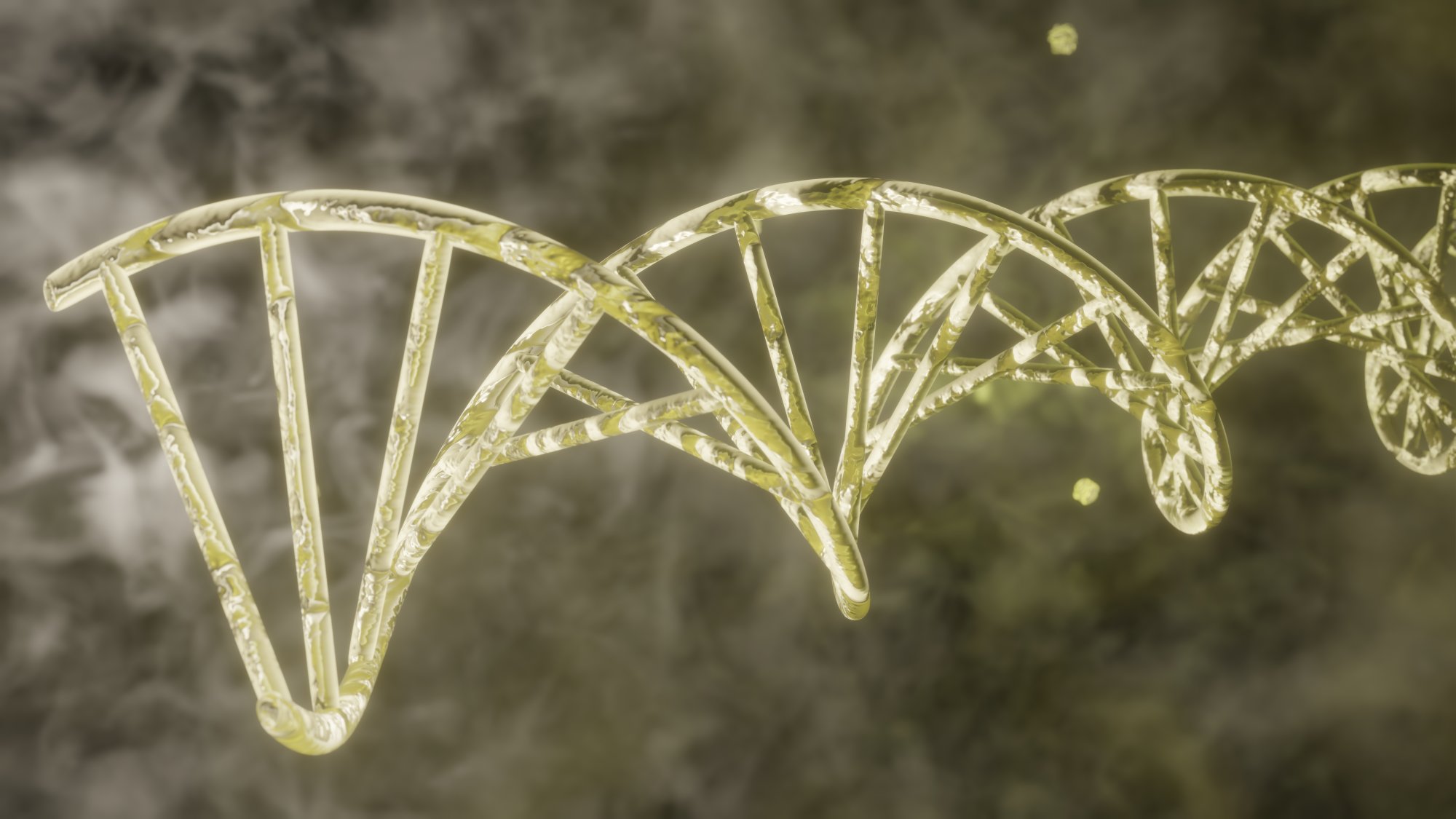
The background environment in the illustration often consists of translucent textures and soft gradients, symbolizing tissue layers, fluid pathways, or cellular spaces. These artistic details provide context, helping viewers imagine how bacterias navigate through biological systems. Subtle lighting effects, reflections, and shadows further enhance realism, making the illustration feel like a live microscopic view.
Additionally, the 3D scene may depict clusters of bacterias to show colony formation. This grouping reflects the way bacterias organize themselves into communities once they begin multiplying. The illustration visually communicates how infectious agents become more potent over time when left unchecked. This visual message supports medical learning, emphasizing the importance of early detection and treatment.
Beyond medical and scientific education, such illustrations hold significant value in communication materials for health awareness. They simplify complex biological processes, making information accessible to students and the general public. The realistic, detailed nature of the render helps bridge the gap between scientific accuracy and visual engagement, providing a tool that can be used in presentations, textbooks, research papers, and digital platforms.
Overall, the 3D rendered microscopic bacterias and infections illustration serves as a powerful medium for explaining microbial life. Its depth, realism, and clarity offer unparalleled insight into how bacterias function, spread, and interact with living systems. Through a well-crafted visual scene, viewers gain a comprehensive understanding of infection mechanisms, making the illustration an indispensable asset in scientific and medical learning.