Anatomy of Teeth — Structure, Layers, and Functional Roles Explained
The anatomy of teeth represents a finely engineered biological design that supports chewing, speech, digestion, and overall oral stability. Understanding tooth structure is essential for appreciating how daily habits, nutrition, and dental care influence long-term oral wellness. Each tooth is made of multiple layers, specialized tissues, and interconnected components that allow it to withstand constant pressure, temperature changes, and exposure to food particles and microorganisms. Exploring these parts in detail helps explain both normal function and the development of dental conditions.
A tooth can be divided broadly into the crown, the visible region above the gum line, and the root, which anchors the tooth securely into the jawbone. The crown has a protective outer coating called enamel, the hardest substance found in the human body. Enamel shields the inner tissues from physical damage and helps resist wear caused by chewing, biting, and grinding. Although incredibly strong, enamel can gradually weaken through acid exposure, improper brushing, or bacterial activity, making its preservation essential.
Beneath the enamel lies the dentin, a supportive layer that forms the bulk of the tooth's internal structure. Dentin is not as hard as enamel, but it is resilient, allowing it to absorb pressure and protect the sensitive core. This layer contains microscopic tubules that connect deeper tissues to outer regions, allowing sensations such as temperature or pressure to be transmitted. When enamel becomes worn or damaged, dentin exposure can lead to sensitivity, highlighting the importance of maintaining healthy outer layers.
At the center of every tooth is the pulp, a soft tissue chamber containing blood vessels, nerves, and connective fibers. The pulp supplies nutrients and keeps the tooth alive. Its nerve pathways enable sensations that help individuals detect heat, cold, and injury. Inflammation or infection within the pulp, often caused by deep decay or trauma, can result in severe pain and may require specialized treatment to preserve tooth function. Surrounding the pulp and dentin is a foundation of mineralized material that supports the root system.
The root portion extends into the jawbone, held firmly in place by the periodontal ligament. This flexible structure allows teeth to withstand chewing forces while maintaining stability. The surrounding bone acts as a supportive framework, adapting over time in response to pressure and movement. The surface of the root is covered by cementum, a calcified layer that helps the ligament fibers attach securely. Damage to this region can affect overall tooth support and lead to loosening or shifting.
The gum line plays a vital role in protecting deeper tissues. Healthy gums form a tight seal around the base of each tooth, preventing bacteria and debris from entering the underlying structures. When gum tissues recede or become inflamed, the exposed regions can lead to sensitivity, increased risk of decay, and potential bone loss. Understanding the connection between gum health and tooth anatomy emphasizes the importance of daily care, such as brushing, flossing, and professional cleanings.
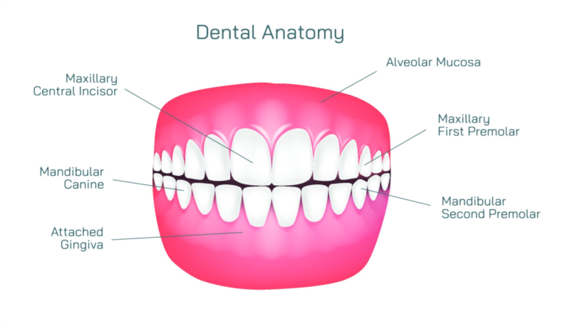
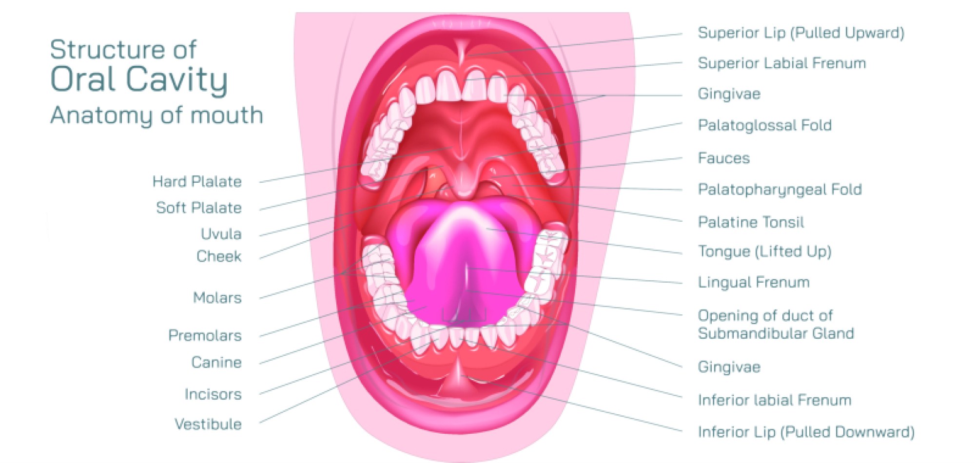
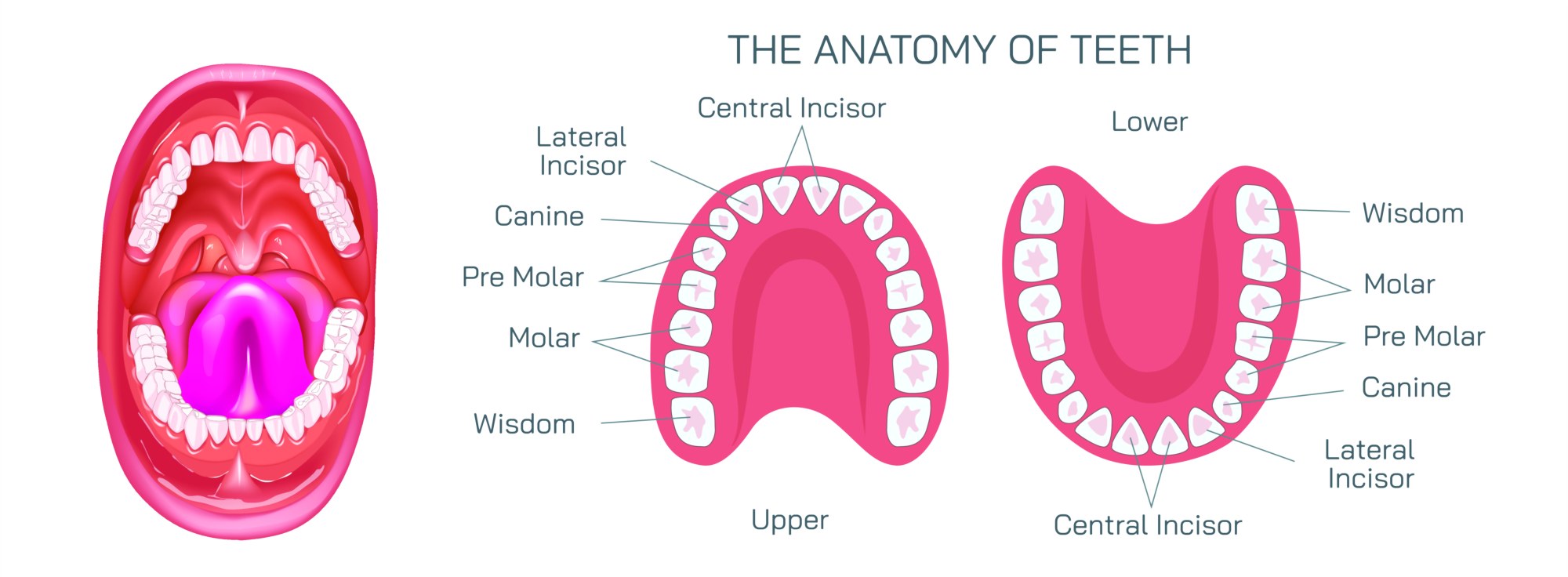
Different tooth types—incisors, canines, premolars, and molars—also reflect structural variations based on their functions. Incisors have sharp edges for cutting food, while canines possess pointed tips for tearing. Premolars combine broad surfaces with ridges suited for crushing, and molars contain multiple cusps designed for grinding. Although their shapes differ, the internal anatomy of each type follows the same layered design, demonstrating a consistent biological pattern adapted for different roles in chewing.
Together, these components create a powerful and efficient system that supports daily oral activity. The anatomy of teeth illustrates how multiple layers work in harmony to protect sensitive tissues, maintain structural strength, and ensure proper dental function. Understanding these features encourages healthier habits, better prevention, and improved oral care decisions that contribute to lifelong dental well-being.