Front Teeth and Molar Teeth — Structure, Function, and Dental Anatomy Explained
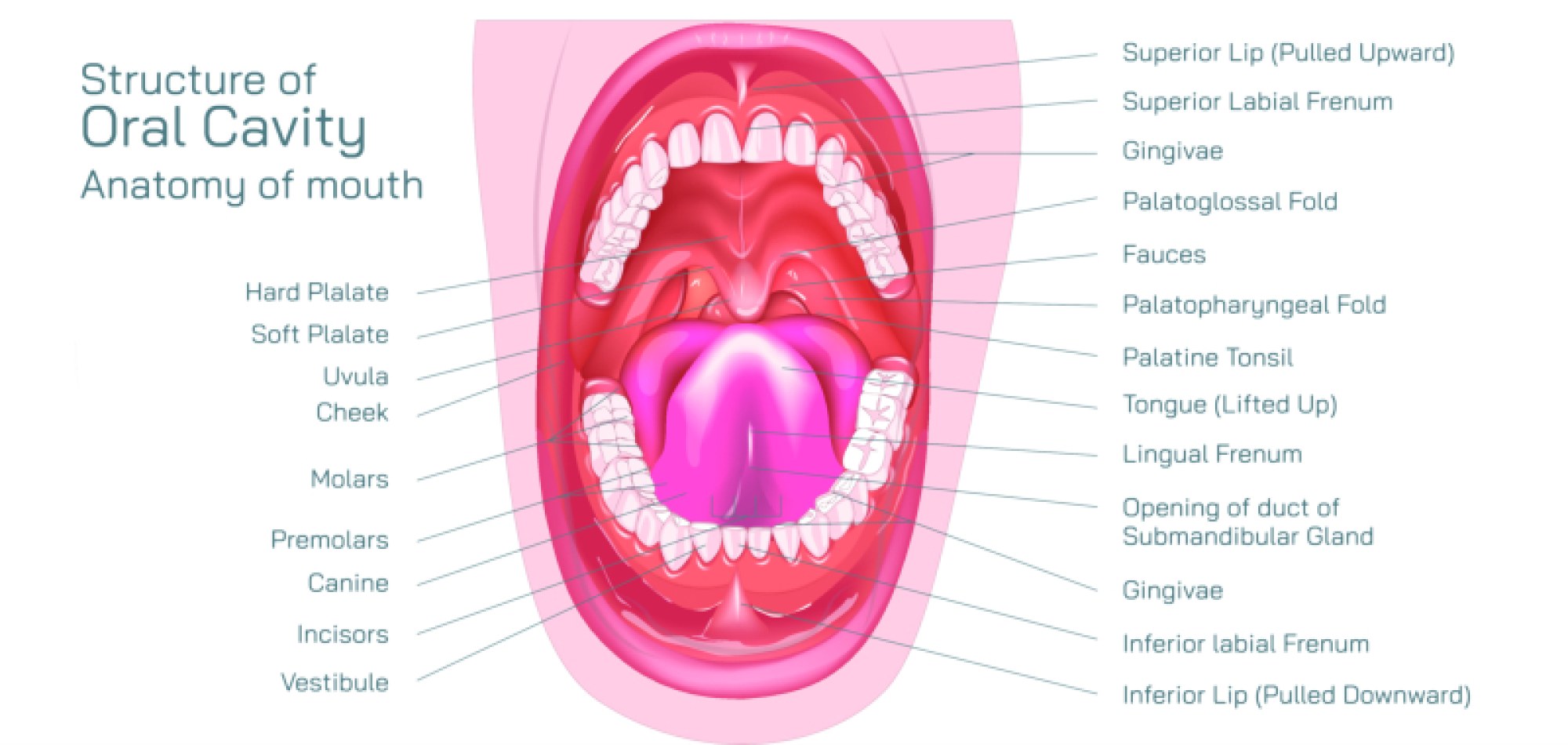
The human mouth is built on a precisely engineered dental system in which every tooth contributes a specialized role to chewing, speech, esthetics, and facial structure. Although all teeth share similar foundational components—enamel, dentin, pulp, cementum, and supporting periodontal tissues—they are not identical in shape or purpose. Among them, front teeth and molar teeth represent two ends of the functional spectrum: one designed for cutting and guiding food into the mouth, the other for crushing and grinding food into particles that the digestive system can process. Together, they form a biomechanical chewing sequence that transforms a single act of eating into a coordinated process of biting, tearing, and pulverizing. Understanding the structure and function of front teeth and molars reveals why both regions must remain healthy for proper oral function and why damage in one area disrupts the entire dental system.
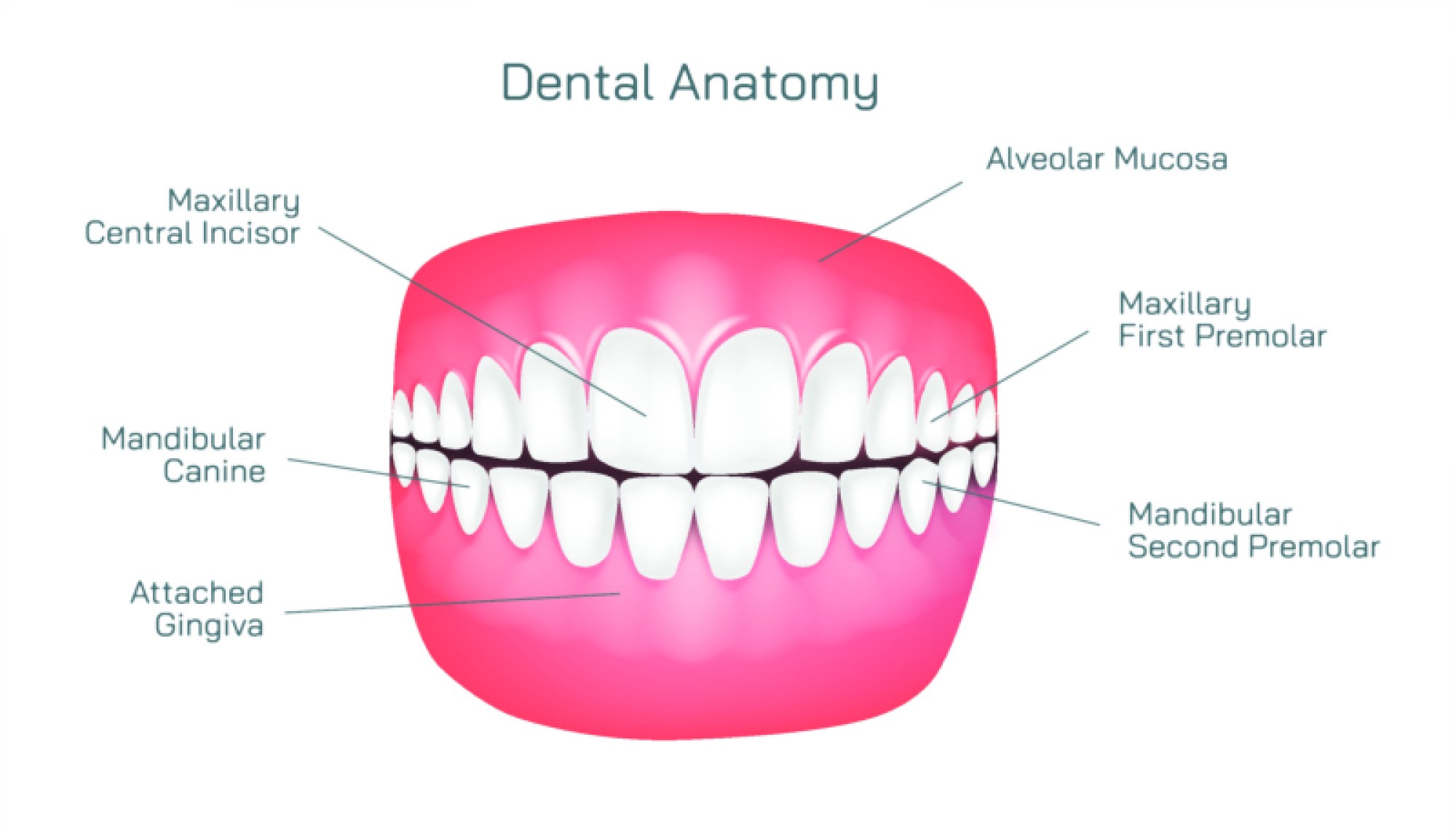
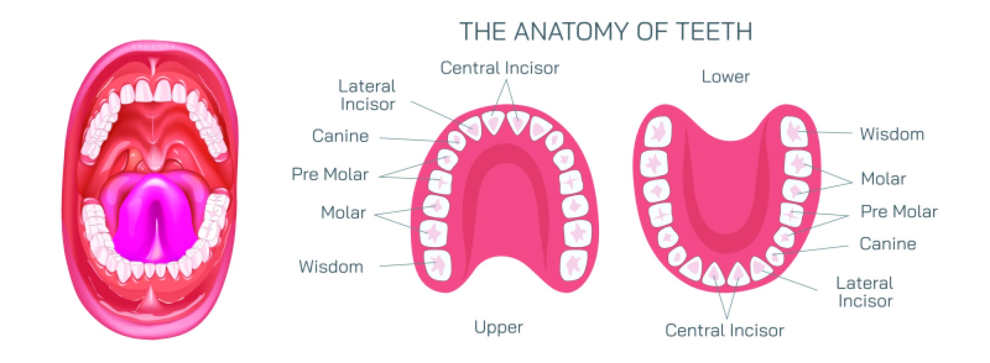
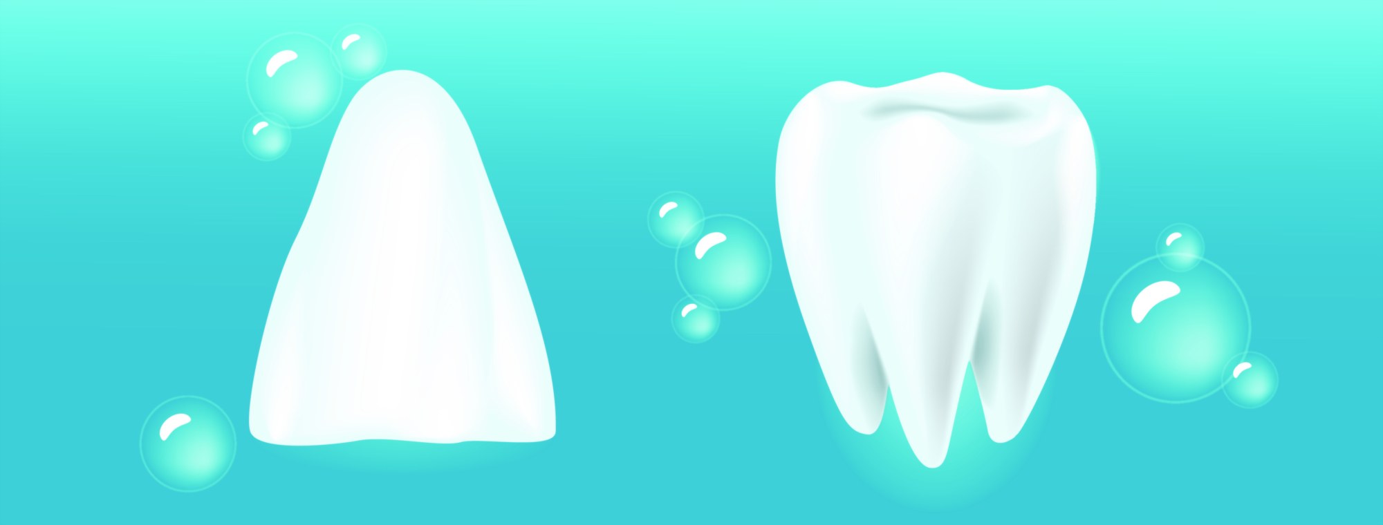
Front teeth consist primarily of incisors and canines, positioned at the visible front of the dental arch. Incisors—the four central teeth and four lateral teeth on the upper and lower jaws—have flat, chisel-shaped edges with sharp cutting surfaces. Their shape allows them to slice food cleanly, acting like blades to break down larger pieces into bite-sized portions. Their design also supports speech production; many sounds require precise contact between the tongue and incisors. Enamel on incisors is slightly thinner than on molars because their primary duty is cutting rather than heavy grinding. Beneath the enamel lies dentin, which surrounds the pulp chamber containing blood vessels and nerves. The roots of incisors are single and straight, enabling stability while maintaining a slim shape that fits neatly in the front of the arch. Because they are constantly exposed during smiling and talking, incisors play a large role in facial expression and appearance. When incisors are misaligned, worn, chipped, or discolored, esthetic changes become immediately visible—demonstrating their emotional and psychological significance as well as their mechanical purpose.
Canines, also located in the front section of the mouth adjacent to the incisors, add an additional layer of function and structure. Their pointed cusp and long root make them ideal for tearing dense food textures, serving as transition teeth between cutting incisors and grinding molars. Canines also play a critical role in guiding the jaw during side-to-side motion, protecting the posterior teeth from damaging friction forces. Their roots are the longest in the mouth, anchoring them firmly and providing reinforcement to the curvature of the dental arch. Because of their strength, canines act as stabilizing “cornerstones” of the mouth—they influence bite alignment, facial support, and orthodontic planning. Together, incisors and canines form the functional and esthetic front unit of the smile, responsible for cutting, tearing, articulation of speech, and shaping the profile of the lips and cheeks.
In contrast, molar teeth are positioned at the back of the mouth and serve as the powerhouse of the chewing system. Molars—12 in total across the adult mouth—have broad, multi-cusped chewing surfaces designed to crush and grind food thoroughly. Their shape reflects their duty: instead of a thin cutting edge, each molar has a wide crown with deep grooves, pits, and fissures to break down food into fine particles that are safe to swallow and easy to digest. Molars endure the highest chewing forces, often several times greater than the pressure exerted on front teeth. To support this mechanical load, molars possess large roots—two or three branches depending on their position—that disperse force into the surrounding jawbone. The thick enamel layer on the occlusal (chewing) surface of molars protects them from wear, although it does not make them immune to decay; their deep grooves easily trap food and plaque, making them especially susceptible to cavities if oral hygiene is insufficient.
Molars are also essential to maintaining vertical facial height—the distance between the upper and lower jaws. When molars are lost or severely worn down, the bite collapses, reducing facial support and contributing to premature wrinkling, jaw soreness, and difficulty chewing. Molars provide symmetry and structural relationship across both sides of the mouth, and losing even one molar can trigger a chain reaction of shifting teeth, misaligned bite, and increased wear on remaining teeth. This demonstrates how the absence of molars affects not only chewing ability but also joint function, muscle balance, and long-term oral health.
When the roles of front teeth and molars are compared, their differences reveal a striking complementarity. Front teeth are precise instruments designed for initiating eating, while molars serve as the final processing station before swallowing. Without front teeth, eating becomes clumsy and inefficient because food must be torn or cut using side teeth or molars, increasing risk of soft tissue injury and excessive pressure on posterior teeth. Without molars, food passes through the mouth insufficiently ground, which affects digestion and forces front teeth to absorb stress they were not designed to handle. A healthy dental system therefore depends on cooperation between the front and back teeth. When they work together, chewing becomes a smooth mechanical cycle: incisors bite, canines tear, premolars start grinding, and molars complete grinding.
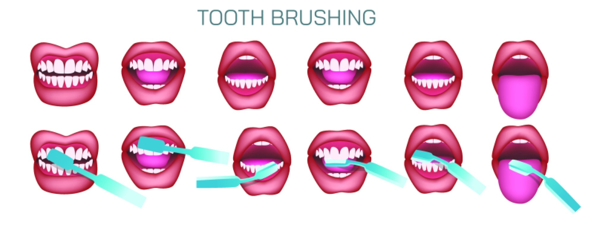
Daily care needs for front teeth and molars reflect their structural differences. Front teeth are vulnerable to erosion and trauma but easy to clean because their surfaces are smooth and accessible. Molars, on the other hand, are more protected inside the mouth yet more vulnerable to decay because brushing and flossing must reach into their deep grooves and contact points. Sealants on molars for children and young adults often serve as a preventive measure, protecting these complex chewing surfaces from bacterial buildup. Grinding and clenching—whether due to stress, sleep disorders, or bite imbalance—tend to affect molars most strongly and may require night guards to prevent long-term wear. Fluoride strengthens enamel across all teeth, but its protective benefit is especially valuable for molars due to their thick chewing surfaces and resistance demands.
Orthodontic alignment connects front and molar teeth into a unified system. When front teeth are properly positioned, they distribute forces evenly and prevent molars from absorbing excessive impact. Molars in correct alignment allow proper jaw movement and ensure that front teeth do not strike prematurely, protecting enamel from fracture and wear. A healthy bite is not judged by esthetics alone but by the balance of forces between front and back teeth—evidence that dental anatomy and function cannot be separated.
Ultimately, the relationship between front teeth and molar teeth defines how the mouth works. Front teeth provide precision, control, and esthetics; molars provide strength, stability, and efficiency. Every meal, every word spoken, and every smile shared reflects their cooperation. Damage or decay in either region affects the whole system because teeth do not function individually but as parts of an interconnected design. Maintaining both front teeth and molars through consistent oral hygiene, balanced diet, fluoride use, flossing, proper brushing technique, and regular dental check-ups preserves not just tooth structure but the overall integrity of the chewing mechanism, facial harmony, and quality of life. When the entire dental arch is protected and functioning smoothly, the result is more than a healthy mouth — it is a confident and lasting smile supported by the full architecture of the human dentition.