Comprehensive Guide to MRI Scan Color Codes and Tissue Signal Interpretation
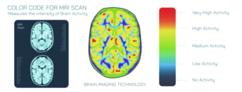
MRI scans use a structured system of shades to represent how tissues respond to magnetic fields and radio waves. These shades do not reflect real colors but instead form signal patterns that help identify structure, density, and internal changes. Understanding these tones allows learners to interpret typical views with greater clarity, making it easier to recognize essential regions and distinguish normal findings from unusual patterns.
Bright areas indicate regions that return strong signals. These zones often correspond to softer structures or areas containing more fluid. Their brightness helps highlight key features, allowing viewers to see contrast between nearby layers. Mid-tone regions carry moderate responses. These areas often represent balanced tissue types that fall between soft and firm consistency. Their neutral appearance helps build an overall structural map across the image.
Dark zones show weaker signal responses. These areas typically correspond to dense regions, flowing spaces, or parts with minimal internal activity. The darker appearance helps define borders, outline shapes, and support recognition of deeper layers. By understanding how these zones interact, learners gain stronger insight into internal patterns and can better follow the natural arrangement of the scanned area.
Together, these tones create a readable layout that guides viewers through each section of the image. The interplay between bright signals, mid-tones, and dark zones forms a complete picture of internal balance, helping students, clinicians, and researchers build skill in reading diagnostic scans. With practice, this understanding improves visual accuracy, supports confident interpretation, and deepens overall familiarity with imaging patterns.