Parts of a Leaf and Leaf Anatomy Vector Illustration Showing Structure, Veins, Stomata, and Photosynthesis Regions
Leaves are the primary organs of plants responsible for photosynthesis, transpiration, and gas exchange, serving as the interface between the plant and its environment. Understanding leaf anatomy is essential for studying plant physiology, ecology, and botany. A vector illustration of a leaf typically highlights its external and internal structure, venation patterns, stomatal distribution, and regions involved in photosynthesis, providing a comprehensive visual guide to its functional and structural components. By integrating anatomical labeling, cross-sectional views, and physiological context, such illustrations enable learners to connect form with function in plant biology.
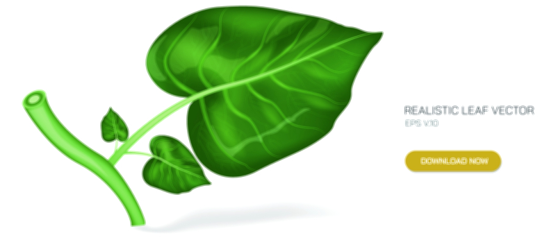
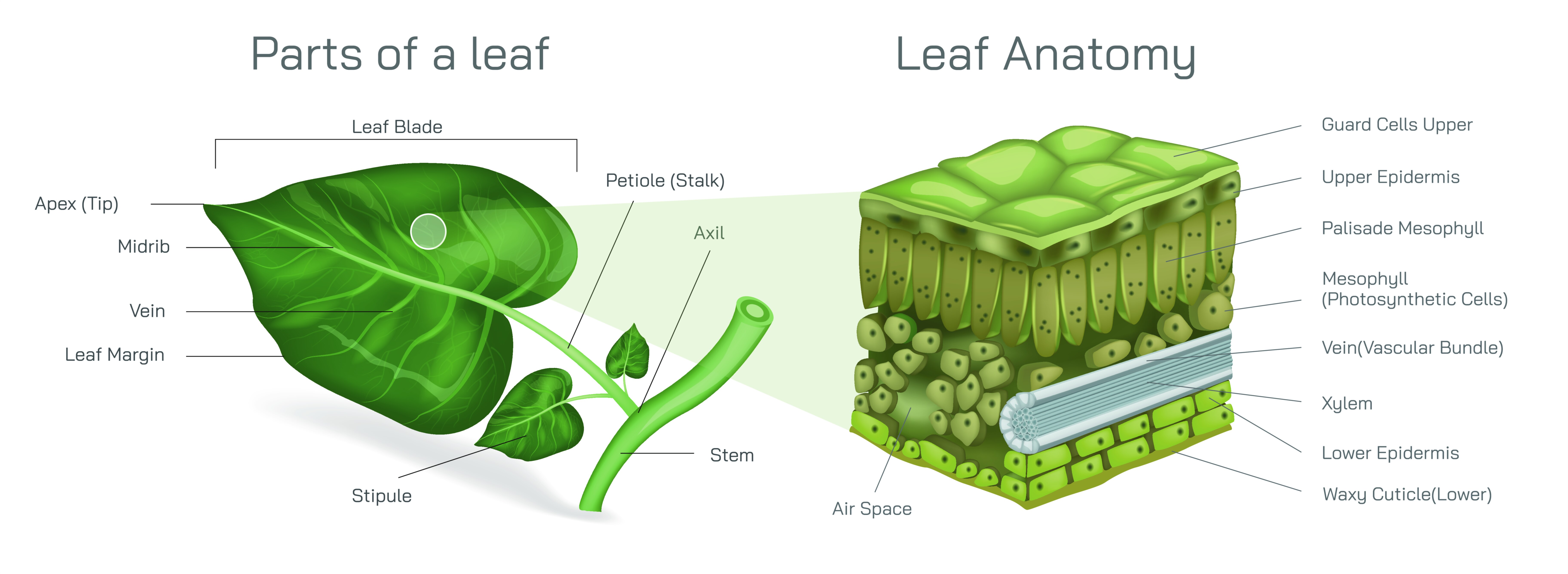
At the most basic level, the external structure of a leaf is depicted, including the leaf blade (lamina), midrib, petiole, and leaf margin. The lamina represents the broad, flattened surface where photosynthesis primarily occurs. The midrib, the central thick vein, provides mechanical support and houses vascular tissues for transport of water, nutrients, and photosynthates. The petiole connects the lamina to the stem, enabling flexibility and positioning for optimal sunlight exposure. In vector diagrams, these structures are often color-coded and labeled, with arrows indicating the orientation of the leaf on the plant and the flow of materials from stem to leaf. Leaf margins may be depicted as entire, serrated, lobed, or dentate, illustrating morphological diversity.
A central feature of leaf illustrations is the venation system, which provides both structural support and transport pathways for water, minerals, and photosynthates. Venation is typically classified as pinnate, with a single midrib and lateral veins, or palmate, with several primary veins radiating from a common point. Secondary and tertiary veins form a network, visible in cross-sectional or transparent views in vector diagrams. Color-coded arrows may indicate the flow of xylem sap from roots to leaf tissues and phloem sap transporting sugars from leaf to stem, demonstrating the dual transport function of the vascular system. The venation pattern also contributes to leaf strength and flexibility, which can be highlighted in schematic overlays or magnified sections.
Internal leaf anatomy is another crucial component. Vector illustrations often include a cross-sectional view of the leaf to show the epidermis, mesophyll, and vascular bundles. The upper and lower epidermis are depicted as protective layers, often covered by a cuticle that reduces water loss. The palisade mesophyll, located beneath the upper epidermis, is shown as tightly packed cells rich in chloroplasts, representing the primary site of photosynthesis. Beneath this layer, the spongy mesophyll contains loosely arranged cells with intercellular air spaces, facilitating gas exchange. Labels and arrows indicate the movement of carbon dioxide into the leaf and oxygen out, as well as the transport of water and nutrients from vascular tissues to photosynthetic cells.
Stomata are specialized pores found primarily in the lower epidermis, regulating gas exchange and transpiration. In vector diagrams, stomata are often magnified to show guard cells, which control pore opening and closing. Arrows may indicate the movement of CO₂ into the leaf for photosynthesis and O₂ and water vapor out during transpiration. The illustration can also show the response of stomata to environmental cues such as light, humidity, and internal water status, emphasizing their dynamic role in leaf physiology. Color coding or shading helps differentiate stomata from surrounding epidermal cells, making their location and function visually clear.
Vector illustrations often highlight the photosynthesis regions within the leaf. Chloroplast-rich palisade mesophyll is the primary site of light-dependent reactions, while spongy mesophyll supports gas diffusion and contains additional chloroplasts for carbon fixation. Arrows may indicate the flow of electrons, water, and carbon dioxide during photosynthesis in simplified form, connecting anatomical structure with biochemical function. The depiction may include labels for chloroplasts, thylakoid regions, and cytoplasmic connections, offering a detailed yet accessible visualization of photosynthetic activity.
Additional features in vector diagrams may include leaf modifications such as tendrils, spines, or succulent leaves, demonstrating how structural variation aligns with ecological adaptation. The illustration may also show vascular bundle magnification, highlighting xylem and phloem components within the leaf and their role in nutrient and water transport. Labels, arrows, and color gradients enhance clarity, showing how the internal structure supports leaf function and overall plant physiology.
By combining external morphology, venation patterns, cross-sectional anatomy, stomatal distribution, and photosynthetic regions, a vector illustration of leaf anatomy provides a holistic understanding of this essential organ. It demonstrates how structural components support physiological processes, including energy capture, gas exchange, water regulation, and nutrient transport. Comparative panels may show differences in leaf type, thickness, or venation patterns, further emphasizing functional adaptation.
Ultimately, a vector illustration of leaf anatomy conveys the integration of structure and function, illustrating how leaves are optimized for photosynthesis, transpiration, and plant survival. Through labeled components, cross-sectional views, directional arrows, and color coding, the diagram transforms complex botanical concepts into an intuitive and visually engaging educational tool, enhancing comprehension for students, educators, and researchers alike.