Diplacusis Echoica — Auditory Delay Perception Disorder
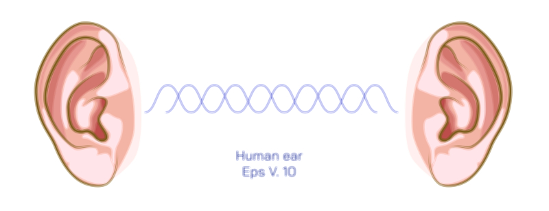
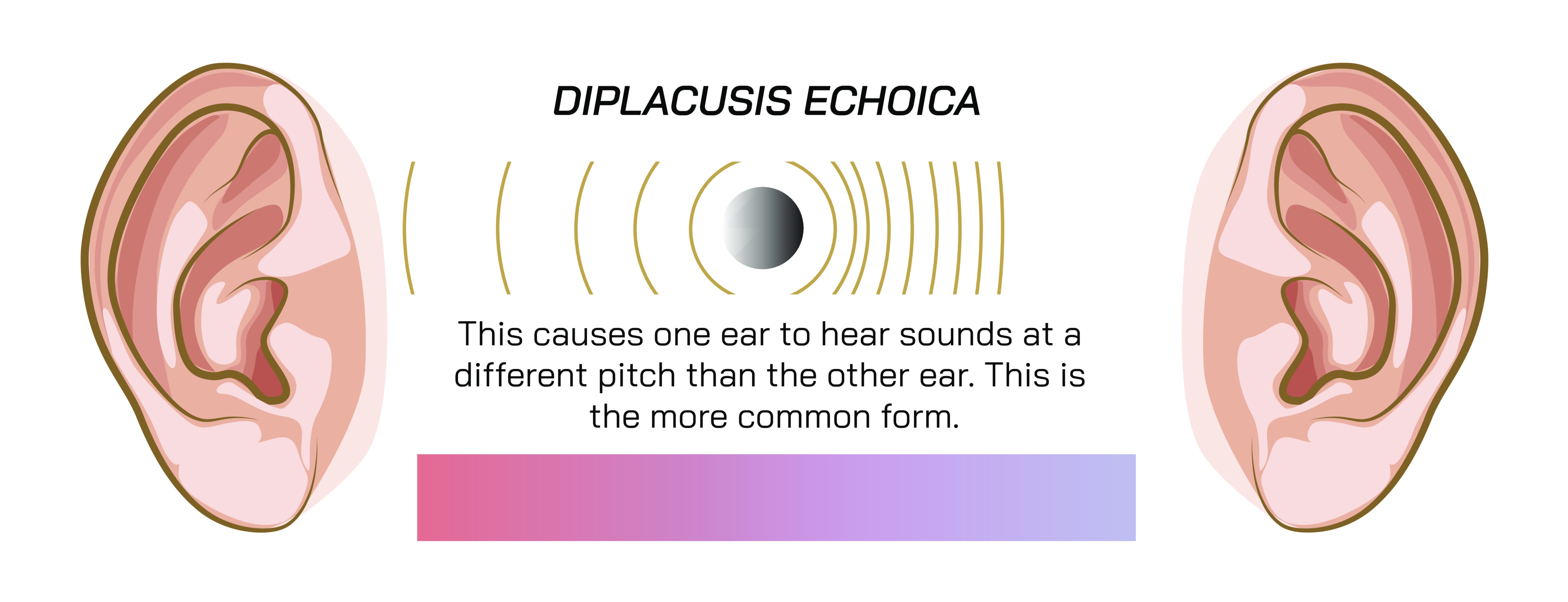
Diplacusis echoica is one of the most unusual and disorienting auditory perception disorders because it disrupts not only the pitch of sound, but the timing with which sound is heard in each ear. Unlike common hearing issues that simply make sounds softer or muffled, diplacusis echoica alters the temporal alignment of auditory information, causing one ear to perceive a sound slightly later than the other. This creates an internal echo or delay, as though a person were hearing the same voice, beep, or musical note twice—once normally, and once with a subtle lag. The condition does not originate from external echoes in the environment; instead, it arises entirely from the way the auditory system processes and transmits sound. Because timing synchronization is essential to forming a unified auditory image, even a tiny delay can disrupt the coherence of sound, leading to confusion, distortion, imbalance, and a persistent sense of auditory dissonance. Diplacusis echoica therefore provides a profound insight into the complex time-sensitive nature of hearing and the importance of synchronized neural processing in both ears.
To understand diplacusis echoica, it is essential to appreciate how the auditory system normally functions. When sound waves enter the ears, they are converted into mechanical vibrations, then fluid waves within the cochlea, and finally electrical signals transmitted along the auditory nerve. These processes happen with astonishing speed and precision—microseconds matter. For the brain to localize sound, interpret pitch, evaluate speech patterns, and perceive timbre clearly, both ears must send synchronized information. Although minor differences in timing between ears are expected—because the ears are physically separated—the brain compensates for natural differences by comparing the signals and constructing a unified perception. Diplacusis echoica occurs when something disrupts this equivalence: one ear may transmit sound signals more slowly due to mechanical dysfunction, neural conduction delays, fluid imbalance, structural damage, or metabolic stress within the cochlea or auditory nerve. This delay forces the brain to treat the incoming information as two separate events, producing the internal echo that defines the disorder.
The echo experienced by individuals with diplacusis echoica can range from an extremely faint, barely perceptible doubling to a pronounced, distracting, or even debilitating auditory distortion. Some describe it as a “shadow” of sound; others hear a distinct repetition, similar to hearing an electronic delay effect applied to speech. In mild cases, the echo might appear only in quiet environments or when listening closely. In more severe cases, every sound—from footsteps to voices to music—seems to split into two, with the delayed version trailing behind. The emotional impact of this can be significant. People may feel disoriented, anxious, or frustrated when their auditory world no longer feels stable or predictable. Musicians, singers, and individuals with strong auditory sensitivity often experience greater distress because even small timing anomalies disrupt their internal sense of rhythm, pitch, and tonal clarity. Diplacusis echoica therefore represents not only a physiological condition but a deeply perceptual one, affecting how people relate to sound and meaning in everyday life.
The causes of diplacusis echoica are varied, but most involve disruptions in the cochlea, auditory nerve, or the neural pathways responsible for synchronizing sound. One of the most common underlying factors is asymmetrical sensorineural hearing loss. When hair cells in one ear are damaged—whether from noise exposure, aging, autoimmune disorders, infections, or vascular issues—the mechanical-to-electrical transduction process becomes inconsistent. Damaged hair cells may respond sluggishly, causing a delay in how the ear translates incoming sound. Fluid buildup or pressure fluctuations in the cochlea, as seen in conditions like Ménière’s disease, can also lead to temporal distortion because changes in endolymph pressure affect how quickly vibrations propagate along the basilar membrane. Middle ear problems such as otitis media or Eustachian tube dysfunction can create mechanical delays by altering the way sound energy reaches the inner ear. Any of these issues can create timing differences between ears that manifest as an internal echo.
Neurological causes can also produce diplacusis echoica. If the auditory nerve is compromised—for example, by demyelination, inflammation, or compression—the electrical signals may travel more slowly from the ear to the brain. Disorders such as multiple sclerosis, acoustic neuroma, or certain neuropathies can produce subtle delays that disrupt the brain’s temporal alignment of auditory information. Even metabolic disturbances affecting neural conduction speed may contribute. In extremely rare cases, central auditory processing disorders or lesions in auditory cortex regions may impair the brain’s ability to synchronize inputs, causing echo-like perceptions even when the ears themselves function normally. These neural causes highlight a fundamental truth about hearing: it is as much a brain-based process as it is an ear-based one, and timing is central to the brain’s ability to fuse sounds into a cohesive whole.
One reason diplacusis echoica is so impactful is that temporal synchronization is essential for speech comprehension. Human speech relies on rapid transitions, short bursts of sound, very specific timing of consonants and vowels, and subtle changes in pitch contour. Even small delays between ears can make speech seem disjointed, metallic, or doubled. This disruption may force the brain to work harder to interpret language, leading to listening fatigue or difficulty concentrating. The person may feel like voices sound “broken,” “split,” or “robotic,” even though no such distortion exists in the external sound environment. Similarly, musical perception suffers greatly because rhythm, harmony, and timbre all depend on unified timing. Instruments may seem out of sync with themselves, or a singer’s voice may appear to reverberate unnaturally, producing discomfort or emotional distress. These perceptual implications underscore the central role of timing in auditory cognition and highlight why even small delays can produce disproportionately large perceptual consequences.
Diagnosis of diplacusis echoica typically begins with a detailed audiological evaluation. Audiologists employ pitch-matching tests, binaural perception assessments, and tone delay recognition exercises to determine whether one ear is transmitting sound slower than the other. Pure-tone audiometry may reveal asymmetrical hearing thresholds, while tympanometry provides insight into middle ear mechanics. Otoacoustic emissions testing can help identify whether specific hair cell regions are malfunctioning. Auditory brainstem response (ABR) tests measure neural conduction timing, making them particularly important when diplacusis echoica appears to have neurological origins. Additional imaging such as MRI may be recommended when acoustic neuroma or other neural pathologies are suspected. Because the condition often coexists with tinnitus, hyperacusis, or sudden hearing loss, a complete diagnostic approach is necessary to distinguish which symptoms arise from which structural or functional changes.
Treatment and management strategies depend on the underlying cause. When diplacusis echoica results from temporary factors such as earwax impaction, middle ear infections, sudden pressure changes, or inflammation, symptoms may resolve once these issues are treated. Addressing sinus congestion or Eustachian tube dysfunction can restore normal middle ear mechanics and eliminate delays. If the cause involves ototoxic medication, adjusting or discontinuing the drug under medical supervision may lead to recovery of normal auditory timing. For cases involving Ménière’s disease or endolymphatic hydrops, controlling fluid pressure through medication, diet, or surgical interventions may reduce the timing mismatch. When sensorineural hearing loss is the underlying cause, hearing aids can be particularly effective because they help rebalance the loudness, clarity, and in some cases timing cues between the ears. Modern digital hearing aids include processing algorithms that align signals across ears, enhancing synchronization. In severe cases involving significant hair cell or neural damage, cochlear implants may offer a pathway to restoring more unified timing.
For some individuals, the brain gradually adapts to the timing mismatch through neuroplasticity. This process can take weeks or months, but the auditory system often learns to reinterpret or suppress the delayed signal. Auditory training exercises—such as sound therapy, temporal discrimination tasks, or binaural alignment activities—may accelerate this adaptation. Musicians sometimes recover better than non-musicians because their auditory pathways are already highly trained in timing and pitch precision, giving them greater neural flexibility. Psychological support, counseling, or coping strategies may also be important because persistent internal echoes can be mentally exhausting or emotionally distressing. Understanding the disorder helps reduce fear or confusion, making the experience easier to manage while recovery unfolds.
Conceptually, diplacusis echoica offers a profound window into the temporal dimension of human hearing. Whereas many auditory disorders affect loudness, clarity, or frequency, diplacusis echoica reveals how deeply we depend on the precise timing of sensory signals to construct a coherent auditory world. The brain is not merely a passive receiver of sound but an active integrator, aligning, comparing, and merging signals from both ears in fractions of milliseconds. When that timing alignment falters, perceptual reality becomes fractured—not because the external world changes, but because the internal processing of sound becomes uneven. In this sense, diplacusis echoica illustrates the elegant fragility of binaural hearing: a process so finely engineered that even tiny delays can produce dramatic perceptual consequences. It also emphasizes the adaptability of the auditory system, which, despite experiencing discord, often works tirelessly to restore coherence.
Ultimately, diplacusis echoica stands as a reminder of the sophistication of hearing itself. It demonstrates how deeply timed, synchronized, and coordinated our auditory perception must be to function seamlessly. It reveals the intricate interplay between mechanical movement in the ear and electrical signaling in the brain. Most importantly, it shows that perception is not simply a reflection of the outside world—it is a precise construction built from the harmony of sensory inputs. When that harmony is disrupted, as in diplacusis echoica, the world can sound doubled, delayed, or distorted, reminding us of the exquisite precision upon which our auditory experience depends.