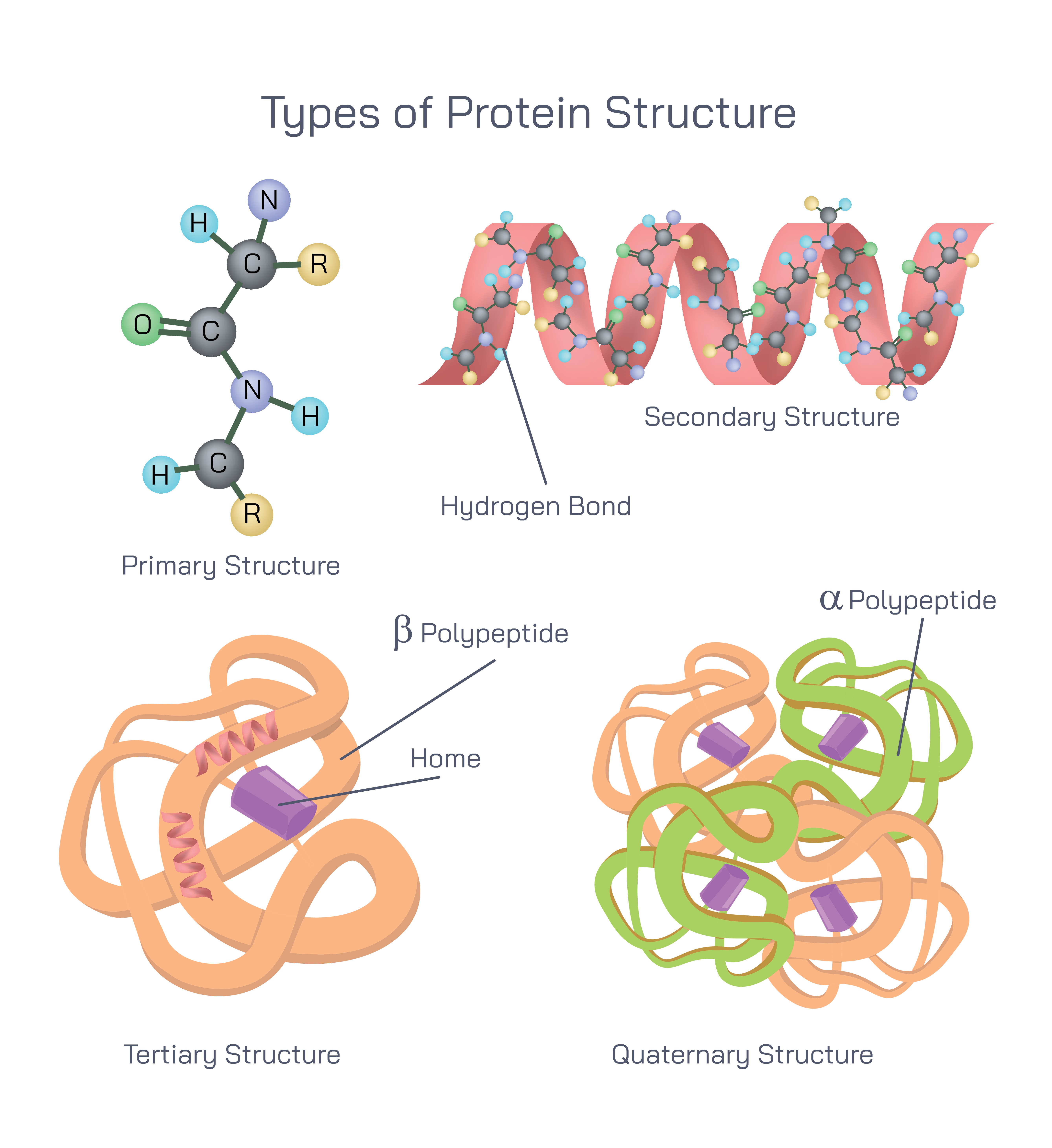
Proteins are essential biological macromolecules that perform a vast range of functions in living organisms, including catalyzing chemical reactions, transporting substances, providing structural support, and regulating cellular processes. The ability of proteins to perform these diverse functions depends entirely on their structure. Protein structure refers to the specific three-dimensional arrangement of atoms within a protein molecule, which is determined by multiple levels of organization. These levels are known as the primary, secondary, tertiary, and quaternary structures of proteins. Each level builds upon the previous one, creating increasing structural complexity and functional specificity.
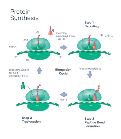
The primary structure of a protein represents the most basic level of protein organization. It consists of a linear sequence of amino acids linked together by peptide bonds. Each amino acid is encoded by genetic information stored in DNA and assembled during protein synthesis at the ribosome. The sequence of amino acids is not random; it is precisely ordered and determines all higher levels of protein structure. Even a small change in the amino acid sequence can significantly alter protein behavior, stability, or function. This sequence acts as the blueprint that guides how the protein will fold and interact with other molecules.
Peptide bonds form when the amino group of one amino acid reacts with the carboxyl group of another, creating a strong covalent bond. This bond links amino acids into a polypeptide chain. The primary structure defines the chemical identity of the protein and establishes the positions of side chains that later interact during folding. Disorders such as sickle cell anemia demonstrate the importance of primary structure, where a single amino acid substitution can lead to dramatic physiological consequences.
The secondary structure of a protein arises from local folding patterns within the polypeptide chain. These patterns are stabilized mainly by hydrogen bonds formed between the backbone atoms of amino acids rather than their side chains. The two most common secondary structures are the alpha helix and the beta sheet. In an alpha helix, the polypeptide chain coils into a spiral shape, with hydrogen bonds forming between nearby segments of the chain. This structure provides flexibility and strength and is commonly found in structural proteins and membrane-associated regions.
Beta sheets form when segments of the polypeptide chain align side by side, creating sheet-like structures stabilized by hydrogen bonds between adjacent strands. These strands may run in the same direction or in opposite directions, forming different sheet arrangements. Secondary structures contribute to the overall stability of the protein and serve as building blocks for more complex folding. The combination and distribution of alpha helices and beta sheets vary among proteins, influencing their physical properties and biological roles.
The tertiary structure represents the complete three-dimensional shape of a single polypeptide chain. At this level, secondary structural elements fold further due to interactions among amino acid side chains. These interactions include hydrogen bonding, ionic attraction, hydrophobic interactions, and disulfide linkages. Hydrophobic side chains tend to cluster together away from water, while hydrophilic side chains orient toward the surrounding aqueous environment. This folding pattern allows the protein to achieve a stable, energetically favorable conformation.
Tertiary structure is crucial because it determines the functional regions of the protein, such as active sites in enzymes or binding sites for other molecules. The precise folding enables proteins to interact selectively with specific substrates, receptors, or other biomolecules. If the tertiary structure is disrupted by factors such as temperature changes, pH imbalance, or chemical agents, the protein may lose its functional shape in a process known as denaturation. This highlights the strong relationship between protein structure and biological activity.
Some proteins consist of a single polypeptide chain and function independently once folded into their tertiary structure. Others require additional structural organization to become fully functional. This leads to the fourth level of protein structure, known as quaternary structure. Quaternary structure refers to the assembly of two or more folded polypeptide subunits into a single functional protein complex. Each subunit may have its own tertiary structure, but the interaction among subunits creates new functional properties.
The quaternary structure is stabilized by the same types of interactions found in tertiary structure, including hydrogen bonds, ionic attractions, and hydrophobic interactions. A classic example of quaternary structure is hemoglobin, a protein responsible for oxygen transport in blood. Hemoglobin consists of four polypeptide subunits that work together to bind and release oxygen efficiently. The cooperative interaction among these subunits allows hemoglobin to respond dynamically to oxygen levels in the body.
The hierarchical nature of protein structure demonstrates how complexity arises from simplicity. A linear amino acid sequence folds into local secondary structures, which then assemble into a three-dimensional tertiary form. In some cases, multiple folded units combine to create a quaternary structure with enhanced functionality. This hierarchical organization ensures that proteins are both stable and adaptable, capable of performing precise biological roles under varying conditions.
Protein structure is intimately linked to protein synthesis and cellular function. Proteins are synthesized as linear chains on ribosomes, but folding begins even before synthesis is complete. Specialized cellular environments and helper molecules assist in proper folding to prevent misfolding and aggregation. Proper protein folding is essential for maintaining cellular health, while misfolded proteins are associated with various diseases.
In the broader context of biology and medicine, understanding protein structure is fundamental to fields such as enzymology, genetics, pharmacology, and biotechnology. Drug design often focuses on targeting specific structural features of proteins, while genetic mutations are analyzed based on how they alter protein structure. Structural biology techniques provide detailed insights into how proteins function at the molecular level.
In conclusion, the types of protein structure—primary, secondary, tertiary, and quaternary—represent successive levels of organization that define a protein’s shape and function. The primary structure provides the amino acid sequence, the secondary structure introduces local folding patterns, the tertiary structure creates the complete three-dimensional form, and the quaternary structure enables complex protein assemblies. Together, these levels explain how proteins achieve their remarkable diversity and precision, making them indispensable components of all living systems.