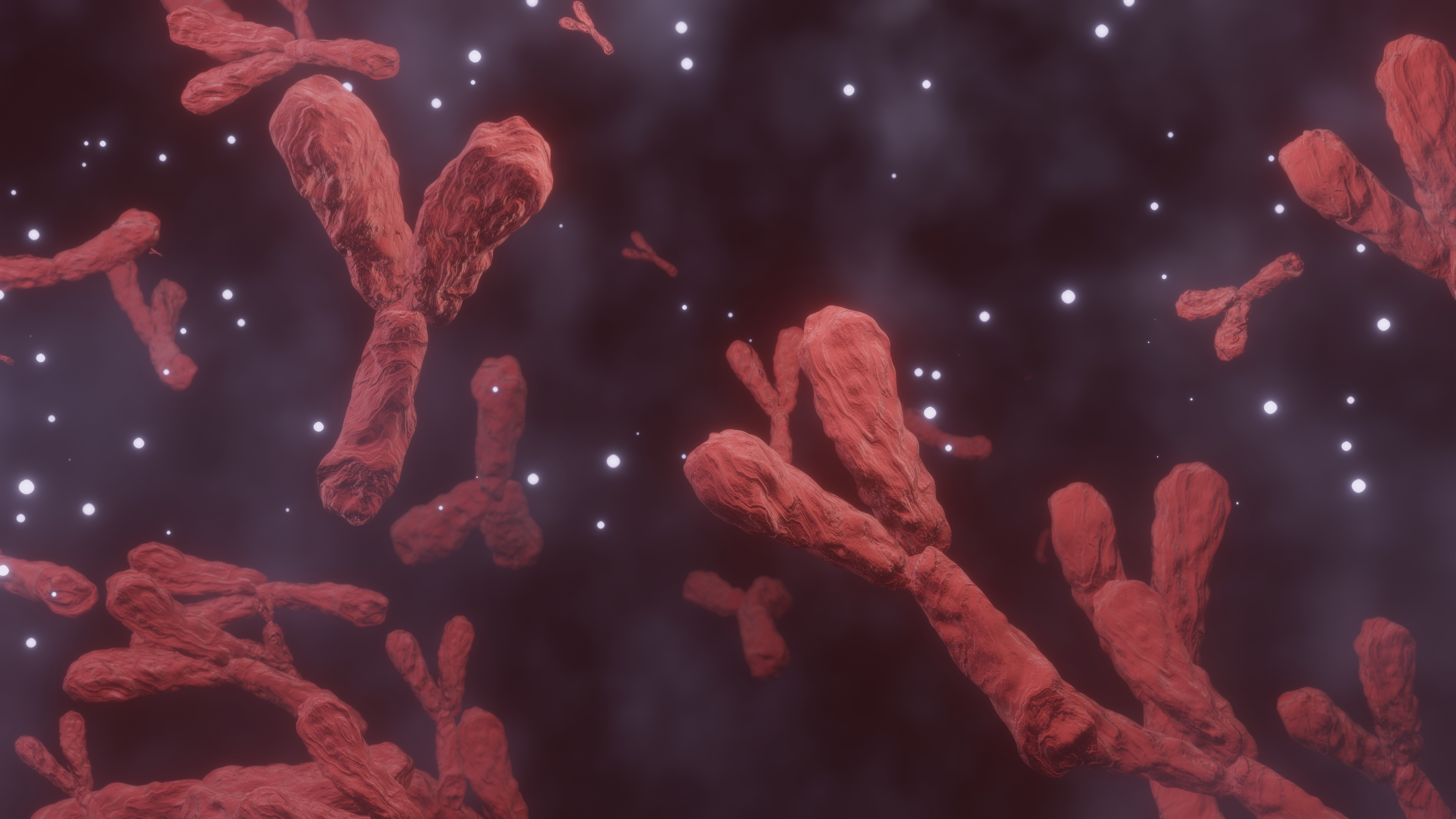
3D Y Chromosome: A Deep Exploration of Its Structure and Biological Significance
The Y chromosome holds a unique place in human biology, not only because of its association with male lineage but also because of its compact, highly specialized nature. When visualized in three dimensions, it becomes far more than a genetic segment; it transforms into an intricate structural landmark within the cell. Observing it in 3D reveals how tightly organized and profoundly functional this chromosome is, despite being one of the smallest in the human genome.
A 3D view shows a slender, curved, condensed form with twisting regions that fold upon themselves with extreme precision. This condensed nature reflects how the chromosome has adapted over countless generations to carry only essential segments required for its primary biological tasks. A close visualization makes the surface appear textured, almost rope like, giving insight into how strands pack together to maintain stability inside the nucleus. This compactness is part of what makes the Y chromosome distinct compared to other larger chromosomes.
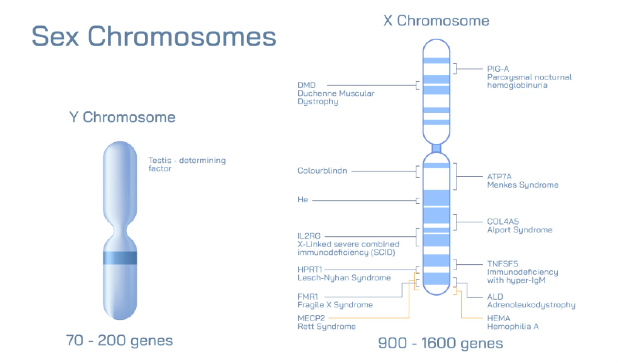
Deep inside its structure lie specific regions responsible for guiding traits related to male characteristics and inheritance. These sections, despite being limited in number, operate with remarkable efficiency. In a 3D model, these areas appear as small organized clusters within the chromosomal body, showing how nature preserves only what is necessary for crucial genetic transmission. The visualization emphasizes the relationship between size and specialization, revealing that the Y chromosome’s influence is not measured by length but by biological focus.
Another significant feature becomes visible when the chromosome is examined from multiple angles. The twisted core resembles a coiled architectural beam, suggesting how its internal support maintains strength even under constant cellular activity. This shape allows the chromosome to survive repeated replication cycles, cellular division, and environmental stress without losing integrity. It offers a striking example of how evolutionary pressure shapes biological structures toward resilience and precision.
The 3D perspective also helps highlight the boundary between the Y chromosome and other genetic components inside the nucleus. Surrounded by numerous larger chromosomes, the Y chromosome stands out due to its distinctive size and shape. Its slender appearance contrasts with neighboring structures, making it easier to identify in scientific imaging. This visual separation reflects its unique origin, evolutionary path, and limited yet essential functional responsibilities.
When scientists analyze a 3D model, they often focus on the organization of chromatin layers within the chromosome. These layers fold into progressively smaller loops that lock into position like an intricately arranged internal framework. This controlled folding allows specific genetic regions to remain accessible when needed and protected when not in use. The visualization helps learners understand how biological systems preserve information in compact spaces while maintaining the ability to retrieve it when signals arise.
One of the most intriguing aspects revealed by 3D observation is how the Y chromosome maintains continuity across generations. It passes almost unchanged from parent to child in the male line, acting as a biological marker of ancestry and identity. This continuity is reflected in its structure, which appears stable, tightly formed, and built to endure through long spans of inheritance. The 3D model makes this endurance more tangible by showing its cohesive design.
A deeper look also reveals variations in density along its length. Some regions appear more compact, suggesting a protective nature, while others seem slightly more open, indicating zones where active biological processes may occur. This variation reflects how different segments serve distinct roles inside the chromosome. Through visualization, these differences become more accessible to learners who might otherwise struggle to understand genetic organization from abstract descriptions.
Visualizing the 3D Y chromosome ultimately enhances understanding by transforming an invisible microscopic element into a detailed, approachable structure. It allows learners to appreciate how biological information can be tightly organized within elegant, functional shapes. The model demonstrates that even the smallest chromosomes carry profound influence through purposeful design, evolutionary refinement, and structural efficiency.
By studying it in this way, one gains deeper appreciation not just for genetics but for the remarkable architecture of life itself. The Y chromosome, though small, stands as a powerful example of how nature balances complexity, precision, and purpose within the tiniest biological spaces.