DNA Structure Showing Double Helix, Base Pairing, and Molecular Genetic Composition in Biology
DNA — deoxyribonucleic acid — is the molecular blueprint of life, carrying the instructions that determine how every living organism grows, functions, and reproduces. Its structure is both elegant and extraordinarily complex, and a vector illustration visualizing DNA typically highlights three fundamental features: the double helix shape, the base pairing system, and the molecular components that encode genetic information. Even though DNA is smaller than microscopic, its architecture explains how life preserves identity from one generation to the next and how billions of cells in the human body share identical instructions.
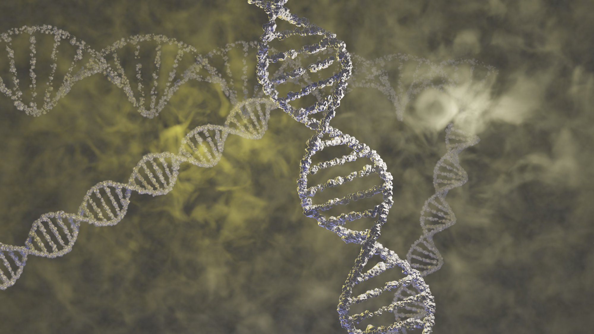
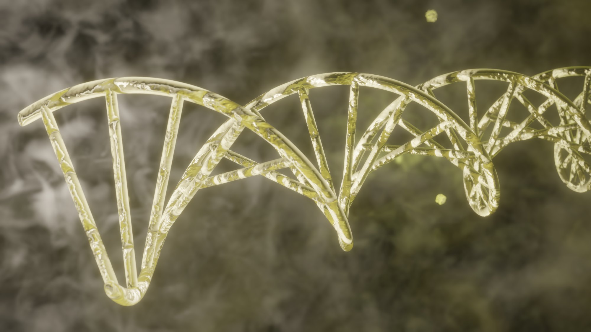
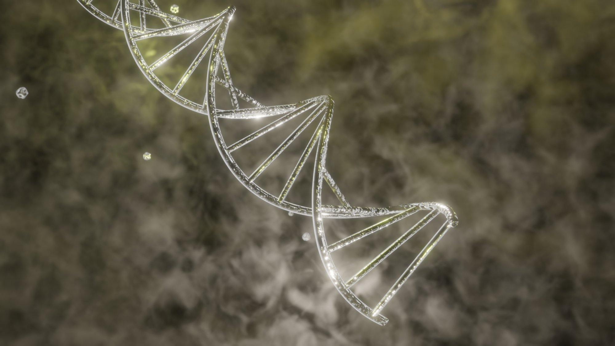
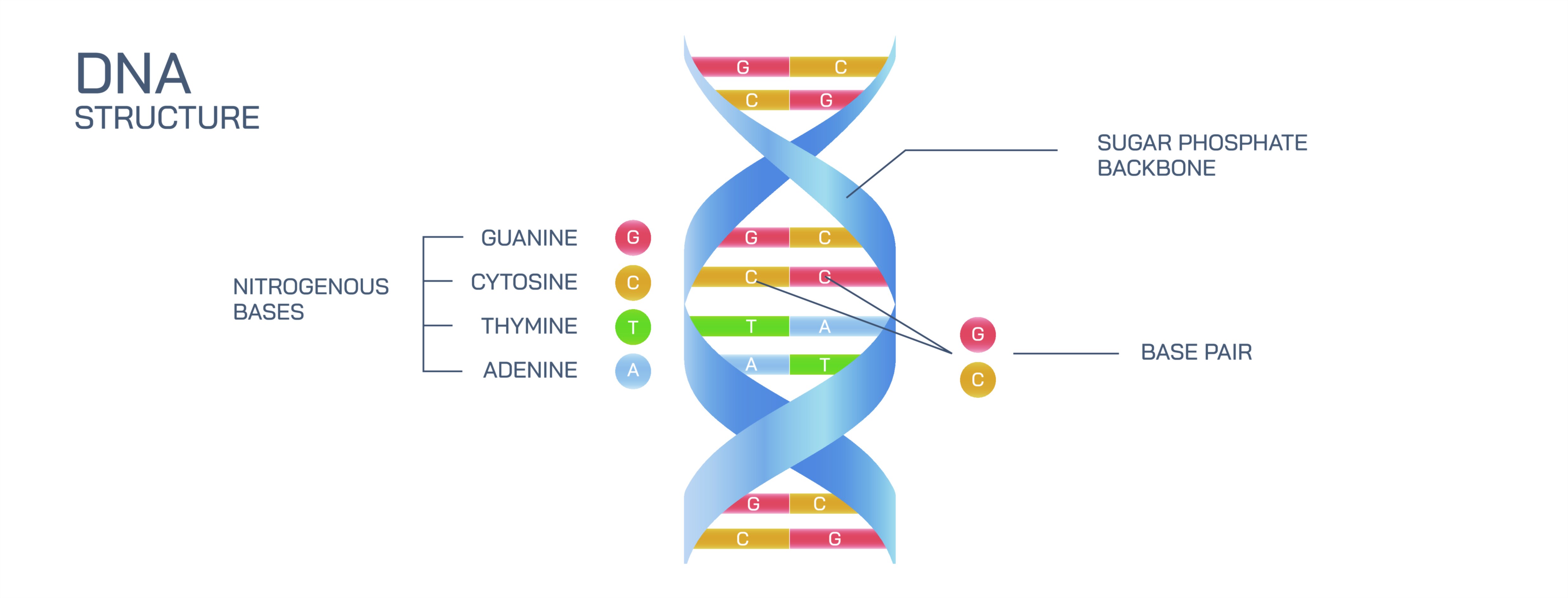
The most iconic feature of DNA is its double helix, a spiral ladder–like structure formed by two long strands twisting around each other. This geometry gives DNA both strength and flexibility. Each strand is made of repeating building blocks called nucleotides, which link end-to-end like beads on a string. In an illustration, these strands appear as two parallel, spiraling rails that wrap around one another in a right-handed twist. The double helix offers stability for storing genetic information yet is easily unwound when the cell needs to copy or read DNA — an essential balance for both continuity and change.
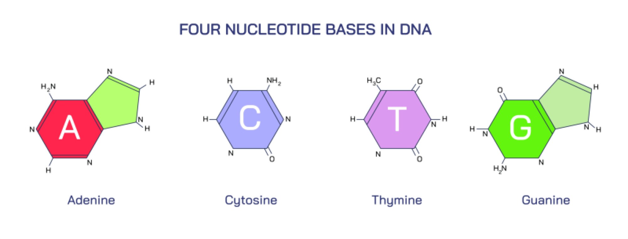
What makes the helix meaningful is the base pairing system, the rungs of the ladder that bridge the two spiraling rails. Each nucleotide contains one of four nitrogen-containing bases:
Adenine (A)
Thymine (T)
Cytosine (C)
Guanine (G)
In the illustration, these bases appear as colored or shaped molecular blocks extending inward from each strand and meeting in the middle to form pairs. The pairing follows strict complementarity: A binds only with T, and C binds only with G. These base pairs are held together by hydrogen bonds — two bonds between A and T, and three between C and G — giving the DNA helix a consistent width and strong but reversible bonding. The base pairing rules guarantee faithful replication because when the two strands separate, each strand carries the exact template needed to rebuild its missing partner.
The backbone of DNA, seen as the vertical sides of the ladder in vector graphics, is composed of alternating sugar (deoxyribose) and phosphate groups. These linkages form a sturdy structural frame, while the bases carry the informational content. The sugars and phosphates connect in opposite directional orientations, meaning the two strands of DNA run antiparallel — one in the 5′→3′ direction and the other 3′→5′. Illustrations often show subtle arrows or labeling to emphasize this polarity, because it explains why enzymes read and copy DNA in specific directions.
The genetic code is stored in the sequential order of the bases along each strand. A long chain of base pairs forms a gene, the unit of DNA that encodes instructions for building a protein. Groups of three bases, called codons, correspond to specific amino acids during protein synthesis. Thus, the base sequence is not just a static pattern but an encoded biological language. In many illustrations, a zoom-in section displays the order of bases (A–T–G–C etc.) to convey this information-rich nature of DNA.
The double-stranded structure also explains how DNA replicates with remarkable accuracy. When a cell prepares to divide, enzymes such as helicase unwind the helix and pull the two strands apart. Each original strand becomes a template for constructing a new complementary strand using the base pairing rules. The result is two DNA molecules, each containing one old strand and one newly synthesized strand — a process called semi-conservative replication. The reliability of base pairing enables inheritance patterns that remain stable while still allowing variation and mutation over evolutionary time.
DNA is tightly packaged inside the cell nucleus. Each molecule wraps around proteins called histones, forming chromatin fibers, which coil further to create chromosomes. This advanced level of organization fits nearly two meters of DNA inside human cells while keeping it accessible for gene expression. Though a full chromosome may not appear in a simple DNA illustration, a connected side diagram often shows DNA coiling progressively into chromatin and chromosomes to provide structural context.
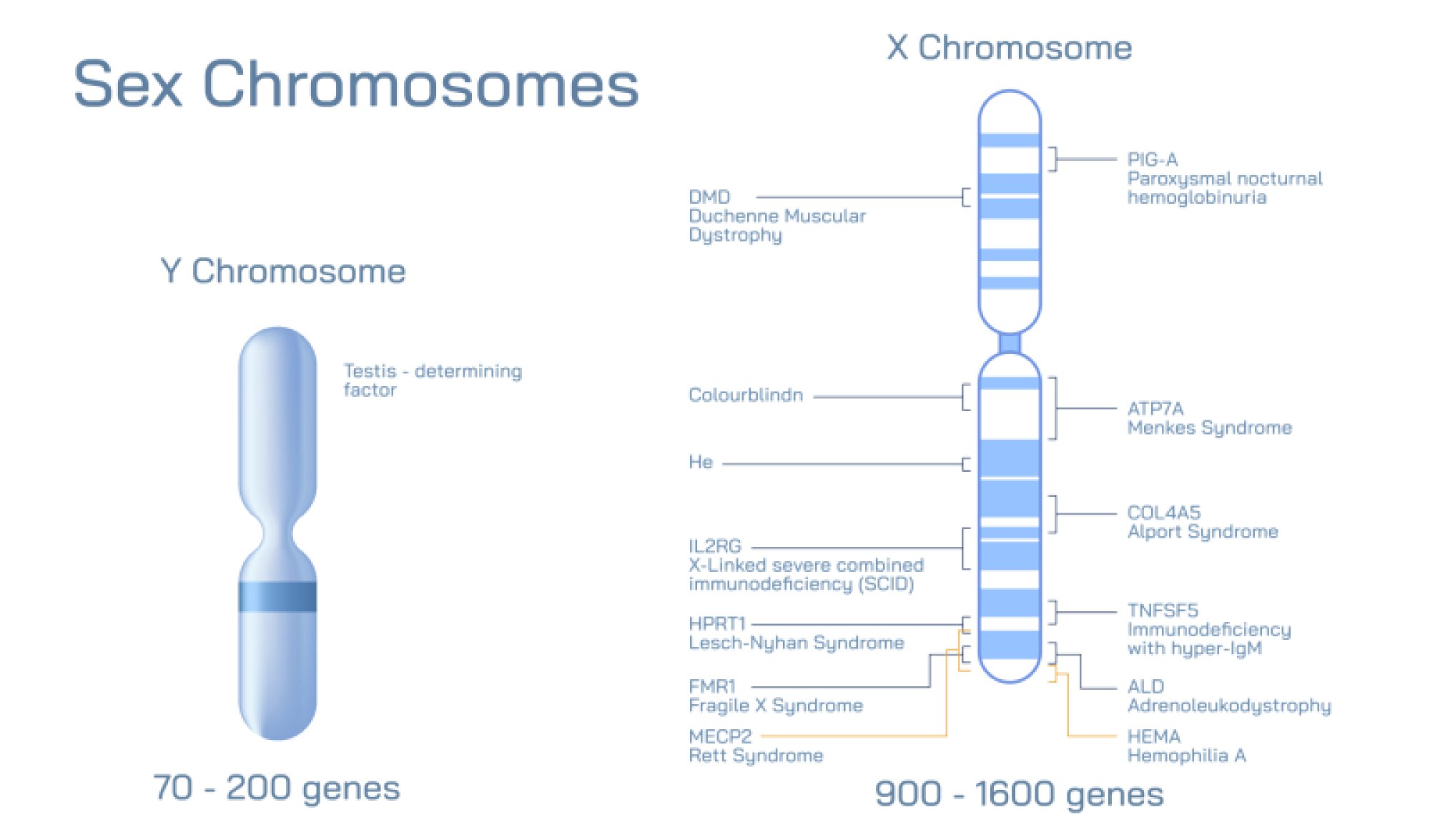
The DNA double helix is also the foundation of biological diversity. Small differences in base sequences distinguish one species from another and one person from the next. Even the slightest change in sequence — a mutation — can alter or impair protein function, sometimes causing genetic disorders and sometimes fueling evolutionary adaptation. This connection explains why DNA analysis underpins medical genetics, ancestry research, forensics, evolutionary biology, and biotechnology.
A complete vector illustration of DNA structure typically includes:
• The double helix, drawn with two twisting strands
• The sugar–phosphate backbone on the outside of each strand
• Base pairs connecting the strands (A–T and C–G)
• Distinct coloring or labeling for each base type
• Optional detail showing the sequence of bases conveying genetic information
• Optional zoom-out graphic showing how DNA folds into chromosomes in the nucleus
Even without chemical formulas or genetic algorithms, this visual representation captures the deep logic of DNA: two strands, held together by precise pairing rules, storing a genome that can be copied, repaired, read, and inherited. The molecule’s structure embodies its function — stability for long-term information storage and flexibility for biological processes.
Through the combined themes of the double helix, base pairing, and molecular genetic composition, the illustration of DNA becomes not only a description of molecular anatomy but a map of how life encodes identity, continuity, evolution, and diversity at the deepest biological level.