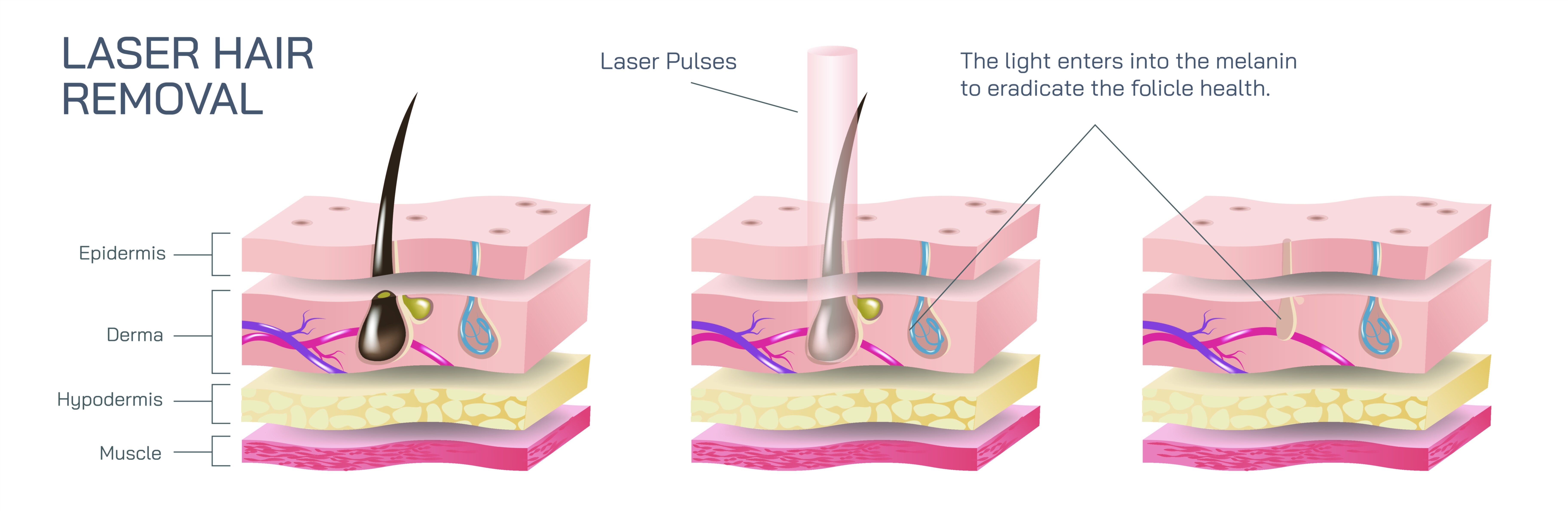
Laser Hair Removal Vector Illustration Showing Skin Layers, Laser Beam Targeting Hair Follicles, and Permanent Hair Reduction Process
Laser hair removal is a widely used cosmetic and dermatological procedure designed to permanently reduce unwanted hair by targeting hair follicles with concentrated light energy. Understanding the underlying mechanism requires knowledge of skin anatomy, hair follicle structure, and laser-tissue interaction. A vector illustration of laser hair removal typically integrates layered skin anatomy, hair follicle targeting, and energy absorption, providing a clear, educational representation of the procedure. By combining labeled skin layers, directional laser beams, and annotations of the hair growth cycle, such diagrams allow learners to visualize both the process and biological rationale behind permanent hair reduction.
At the center of the illustration is a cross-sectional view of human skin, showing the three primary layers: epidermis, dermis, and subcutaneous tissue (hypodermis). Color gradients or shading highlight these layers for visual clarity. The epidermis is labeled as the outermost protective layer, the dermis is shown with embedded hair follicles, sebaceous glands, and connective tissue, while the subcutaneous layer is depicted with adipose tissue and vascular networks. Arrows may indicate the depth of laser penetration relative to skin thickness, emphasizing precise targeting for effective treatment.
Hair follicles are illustrated as tubular structures extending from the epidermal surface down into the dermis. Each follicle includes the hair shaft, follicle bulb, and dermal papilla, with labels identifying the actively growing cells responsible for hair production. Magnified insets may show melanin concentration in the follicle, highlighting the chromophore that absorbs laser energy. Arrows illustrate the laser beam’s path, converging on the follicle bulb where energy absorption triggers selective photothermolysis, the process that damages hair-producing cells while sparing surrounding tissue.
The laser beam is depicted as a directed, narrow light stream entering the skin at the surface and penetrating to the depth of the hair follicle. Color coding—such as red or near-infrared gradients—illustrates energy intensity. Arrows and labels indicate that the beam targets melanin within the follicle, generating heat that destroys the hair root and inhibits regrowth. Additional annotations may explain parameters such as wavelength, pulse duration, and energy density, demonstrating why laser settings must be customized for skin type and hair color.
Vector illustrations often include before-and-after comparisons. For example, an untreated follicle may be shown actively producing hair, while a post-treatment follicle is depicted with damaged bulb cells and reduced growth potential. Arrows may indicate hair miniaturization over successive treatments, visually explaining the cumulative effect of laser sessions. Color-coded overlays can highlight damaged follicles versus unaffected surrounding skin, reinforcing the selective and precise nature of laser therapy.
Additional educational elements include safety and skin protection features. Insets may show the epidermis remaining unharmed due to cooling techniques or absorption specificity, illustrating how the procedure minimizes side effects. Arrows may indicate protective mechanisms, such as skin cooling gels or contact cooling tips, that prevent thermal damage to superficial layers while ensuring adequate energy delivery to hair follicles.
The illustration may also depict the hair growth cycle—anagen (growth), catagen (regression), and telogen (resting)—highlighting that laser treatment is most effective during the anagen phase. Color-coded timelines or arrows indicate follicle activity, explaining why multiple treatment sessions are necessary to target follicles entering the growth phase at different times. Labels clarify that dormant or resting follicles are less responsive, linking treatment planning to biological mechanisms.
By combining layered skin cross-section, labeled hair follicles, laser beam trajectory, selective follicle targeting, and hair growth cycle annotations, a vector illustration provides a comprehensive understanding of laser hair removal. Color coding, directional arrows, magnified insets, and before-and-after panels allow learners to visualize both the anatomical and functional aspects of the procedure, making complex laser-tissue interactions accessible.
Ultimately, a vector illustration of laser hair removal demonstrates the precise mechanism of permanent hair reduction, showing how energy absorption in hair follicles leads to long-term minimization of hair growth while protecting surrounding skin. Through labeled skin layers, hair follicle structures, laser beam paths, and treatment effects, the diagram transforms abstract dermatological concepts into an educational, visually engaging, and intuitive tool for medical students, cosmetic practitioners, and patient education.