3D Insight Into Leg Sensation and Cramp Response — Muscle Signals, Nervous Pathways, and the Physiology of Sudden Contractions
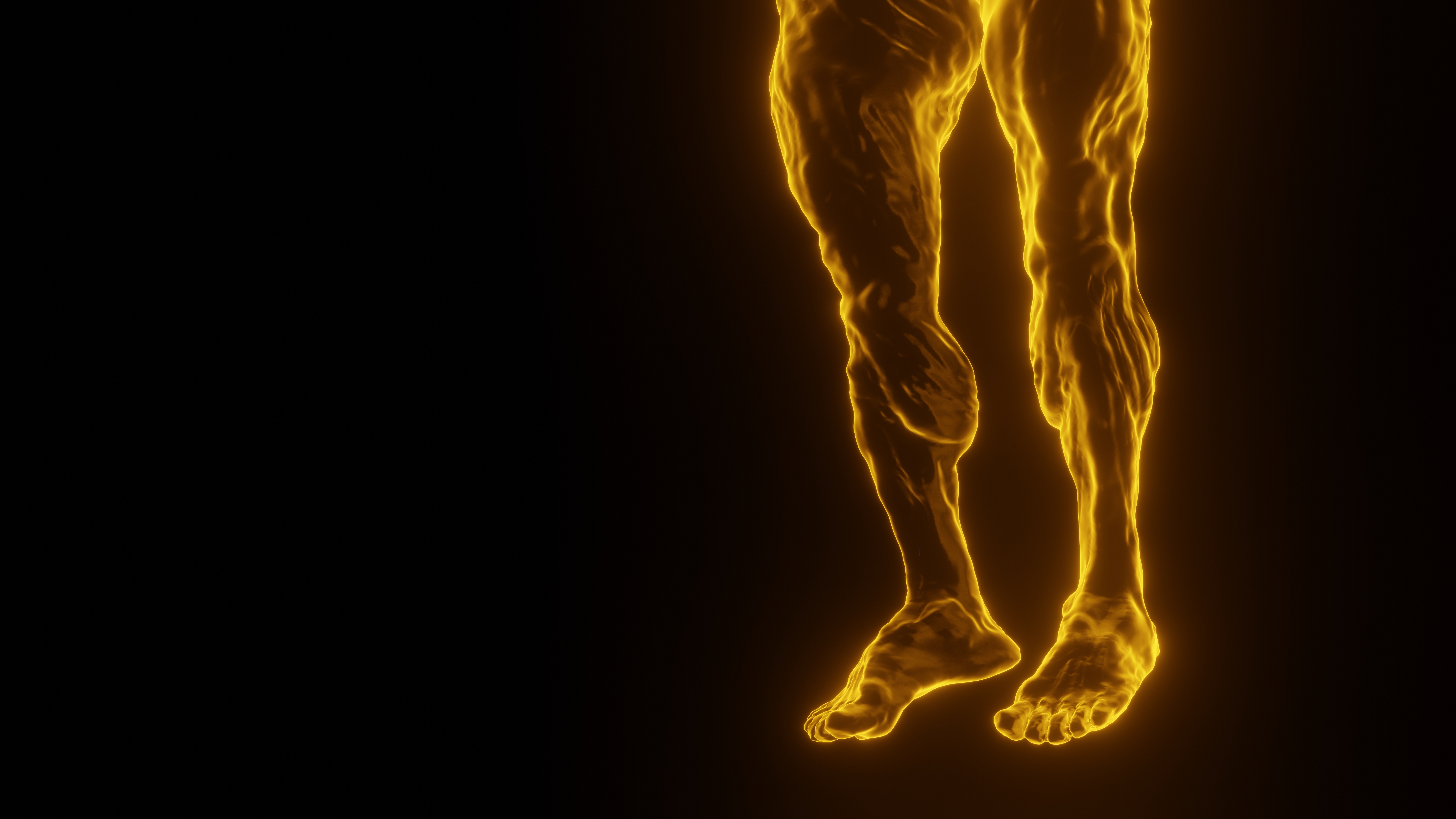
A leg cramp is one of the most striking sensations the human body can generate, creating an abrupt tightening of muscle fibers that feels involuntary, intense, and nearly freezing in place. When viewed through a 3D anatomical and physiological lens, what appears from the outside to be a simple spasm actually represents a complex chain of interconnected events involving motor neurons, sensory nerves, spinal feedback loops, muscle fibers, circulatory supply, electrolyte balance, and biochemical signaling inside the cells. The relationship between sensation and response is so tightly interwoven that the cramp does not simply occur in the muscle alone; it is co-created by the nervous system, the muscle tissue structure, and the communication pathways that regulate contraction and relaxation. When a 3D visualization illustrates the cramp response, it allows us to appreciate the coordinated action of cellular mechanisms, blood vessel behavior, neural reflexes, and sensory perception that together produce the painful tightening and the sudden urge to stretch or massage the leg.
The process begins at the microscopic level inside the muscle fibers themselves, long before pain becomes noticeable. Leg muscles are composed of bundles of fibers that receive instructions from motor neurons, the nerve cells responsible for initiating contraction. These neurons release electrical impulses, or action potentials, which travel down nerve branches into neuromuscular junctions — specialized communication points where chemical messengers called neurotransmitters stimulate muscle fibers to shorten. Under normal movement, this system operates smoothly. The muscle contracts when needed, relaxes when the signal stops, and performs coordinated tasks such as walking, running, or holding a position. However, in a situation that triggers a cramp, the electrical impulses become excessively rapid, unstable, or prolonged, causing the muscle fibers to remain contracted rather than releasing. The muscle cannot lengthen properly because the normal instructions that tell it to relax are either interrupted or overwhelmed. A 3D exploration of the muscle structure at this stage shows tightly packed fiber bundles with cross-bridge attachments locked in place, unable to disengage quickly enough to stop the contraction.
The nervous system plays an equally powerful role in the intensity of leg cramps. Sensory nerves continuously monitor muscle length and tension and send this information to the spinal cord and brain. Under ordinary circumstances, stretch receptors inside the muscle — especially the muscle spindles and the Golgi tendon organs — act as safety regulators. When a muscle contracts, the Golgi tendon organs sense excessive tension and send inhibitory signals that reduce contraction strength to prevent injury. Meanwhile, when a muscle lengthens too fast, muscle spindles signal the nervous system to tighten the muscle slightly to keep movement safe. During a cramp, these protective feedback mechanisms malfunction. The spinal cord reflex arc can amplify contraction rather than moderating it, creating a loop in which the nervous system encourages even more contraction instead of producing relief. A 3D rendering of the nerve pathways highlights how the motor neurons and sensory neurons communicate constantly through branching networks surrounding the muscle, while the spinal cord integrates and reacts to that information instantaneously. When the system becomes overstimulated — because of fatigue, dehydration, mineral imbalance, or prolonged static posture — it can tip into a prolonged contraction without the usual release cycle.
Circulatory factors add another crucial layer to the experience. Muscles depend on steady blood flow to deliver oxygen and electrolytes and remove metabolic by-products generated during physical work. Electrolytes such as calcium, sodium, potassium, and magnesium regulate muscle contraction by controlling the movement of charged particles across cell membranes. When these minerals are imbalanced, the electrical signals that determine contraction and relaxation become erratic. Dehydration, excessive sweating, low blood flow, or sudden exertion can reduce the availability of these electrolytes at the muscle site, making signaling unstable. A 3D view that incorporates blood vessels shows arteries delivering nutrient-rich blood into the muscle before passing through a fine network of capillaries. When fatigue or compression restricts blood flow, waste products like lactic acid accumulate and interfere with proper muscle communication. This biochemical interference contributes to the runaway contraction and heightens the burning discomfort that accompanies a cramp.
The sensation of pain itself is generated through specialized nerve endings embedded throughout the muscle tissue. These nociceptors detect tissue stress, metabolite buildup, and mechanical deformation and then produce sharp, urgent signals that travel rapidly along sensory neurons toward the brain. Pain is not just a side effect; it acts as a biological alarm that forces the person to stop activity and restore normal muscle length. When a cramp occurs, the sensation is often described as gripping, stabbing, or seizing, reflecting how nociceptors fire intensely and without rhythm. A 3D perspective highlights sensory nerves running parallel to motor nerves, demonstrating how sensation accompanies motion rather than being a separate system. This simultaneous flow of information explains why the moment of cramp onset is vivid and overwhelming, especially at night or during exercise when attention is focused elsewhere.
Stretching or massaging the cramping muscle brings relief because these physical actions send strong signals through muscle spindles and tendons that override the faulty reflex arc causing the contraction. Stretching activates receptors that tell the spinal cord to release tension, while massage stimulates circulation and disrupts the locked neural feedback loop. It is not simply the mechanical lengthening of muscle that helps, but the rebalancing of sensory and motor signaling. A 3D sequence of the recovery phase shows the muscle fibers gradually transitioning from rigid cross-bridge locking toward smoother actin–myosin separation, guided by normalized neural impulses and improved blood flow. The sensation of relief is accompanied by a dull soreness because the fibers and surrounding tissue experienced sudden stress during the cramp.
The causes of leg cramps can be diverse, and each root cause corresponds to disruptions in one or more of the systems shown in a deep anatomical visualization. Exercise-induced cramps typically occur when muscle fibers have been repeatedly firing and have reached a point of fatigue where electrolyte reserves and blood flow no longer support smooth contraction. Night cramps may occur when prolonged stillness shortens muscles slightly and alters neural feedback signals. Pregnancy-related cramps involve shifting mineral needs and circulatory adjustments. Cramps in older adults are often linked to age-related changes in nerve conduction, muscle mass, and hydration. In each case, the outward experience is similar — sudden tightening and pain — but the physiological mechanisms driving them differ subtly. A 3D animation helps make these distinctions clearer by visually comparing the various pathways that may be overstimulated or under-supported.
The interaction between the nervous system and the muscles during a cramp also teaches a broader lesson about movement and sensation: the body is constantly balancing contraction and relaxation, activation and inhibition. Muscles are not designed simply to tighten on command; they are regulated by a complex push–pull system of electrical and chemical signals that keeps movement smooth and controlled. When that balance is lost, the body reacts intensely because uncontrolled contraction could damage tissues if it continued unchecked. The cramp acts as both a warning and a self-correcting mechanism, forcing stillness until the nervous system regains control.
Future developments in scientific visualization increasingly use 3D modeling to teach physiology, showing real-time neural firing patterns, blood flow changes, electrolyte flux, and fiber-level contraction. When students and medical professionals see cramps illustrated in dynamic 3D form — not simply as cartoon diagrams but as layered biological processes — they gain deeper appreciation for the coordination required for every simple step, stretch, or posture. The event that feels like a brief moment of discomfort is actually a window into the intricate engineering of the human neuromuscular network.
Understanding leg sensation and cramp responses from a three-dimensional anatomical perspective reveals that the dramatic pain and stiffness are not random malfunctions but rapid protective reactions that emerge from the constant dialogue between muscle cells and the nervous system. The experience of a cramp reflects the body’s extraordinary sensitivity to electrolyte balance, circulation, posture, temperature, and fatigue. It demonstrates how deeply interconnected the brain, spinal cord, nerves, and muscles are, and how the harmony of these systems is essential for smooth movement. What begins as a simple tightening becomes a demonstration of a sophisticated biological process that relies on electrical impulses, molecular signaling, mechanical responses, and pain feedback working in concert. When examined through 3D insight, a leg cramp transforms from a moment of discomfort into a clear example of the human body’s remarkable complexity and the delicate balance that enables effortless movement throughout daily life.