3D Y Chromosome: A Deep Exploration of Its Structure and Biological Significance
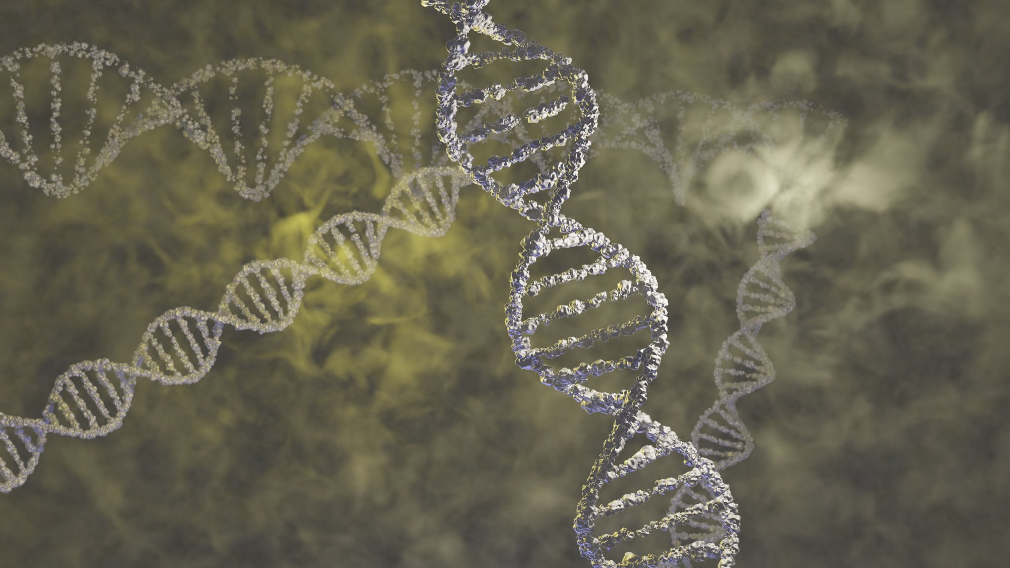
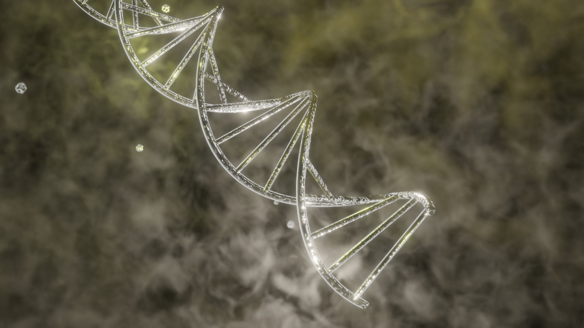
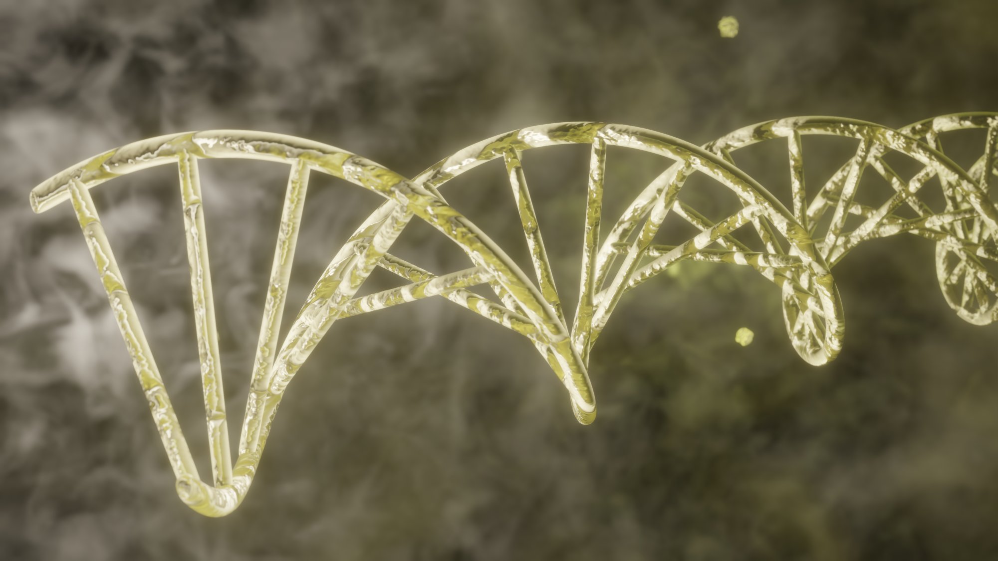
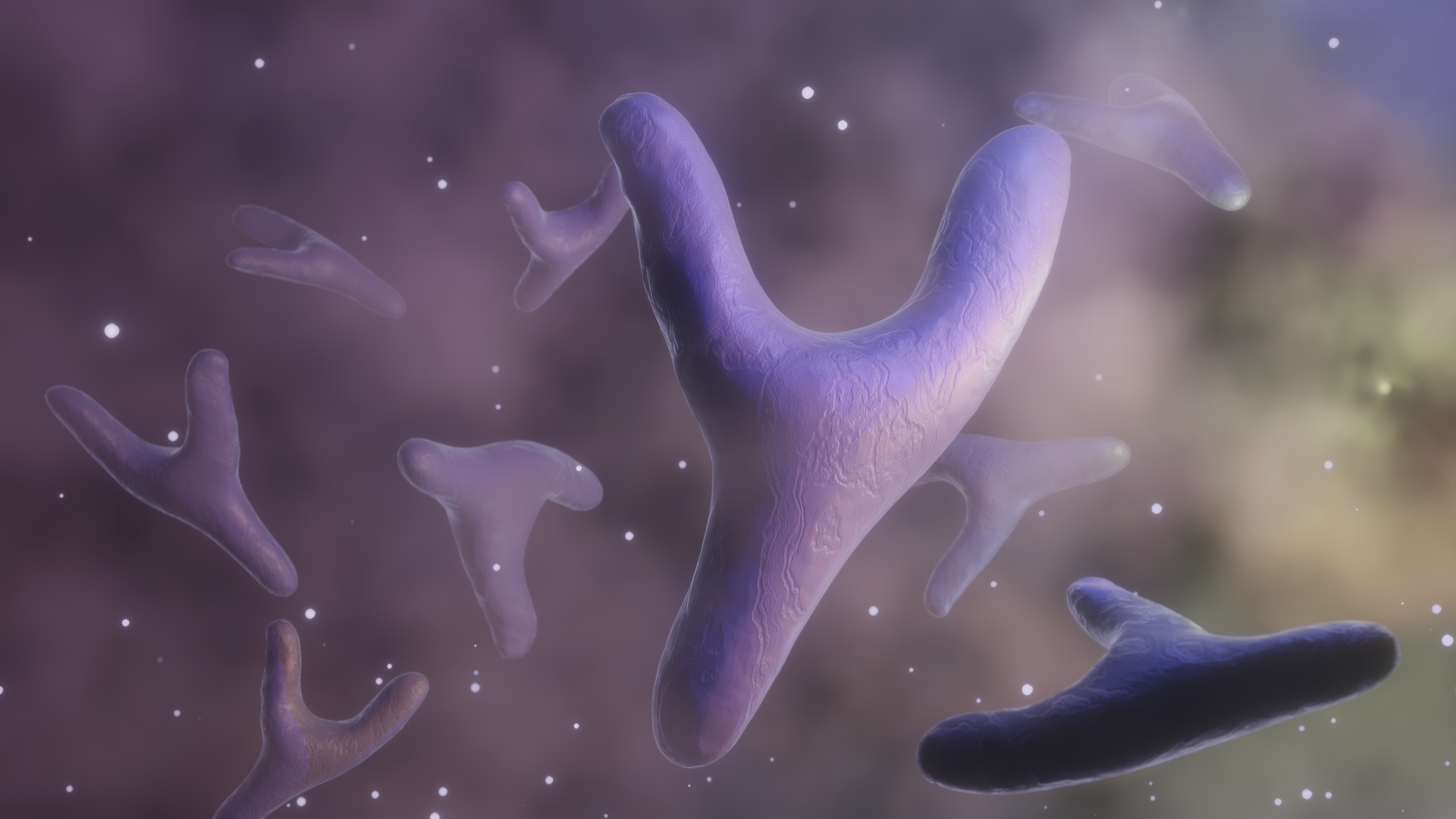
A three dimensional representation of the Y chromosome reveals a remarkable world hidden within the human nucleus. Although it is one of the smallest chromosomes, its shape, arrangement, and function are profoundly meaningful. When viewed in 3D rather than flat diagrams, the Y chromosome appears as an intricately folded structure, shaped like a compact tower with twisting layers pressed together to create a resilient genetic form. This model allows learners to appreciate its tight organization, its preserved regions, and its focused biological role.
The 3D view brings attention to the way its outer layers create a textured surface. Instead of appearing smooth, it displays subtle strands and grooves that hint at winding chromatin paths beneath. These patterns reveal how the chromosome fits enormous genetic information into a remarkably small physical space. Each fold has purpose. Each twist contributes to protection, accessibility, and internal balance. Seen from different angles, the chromosome resembles an artistic blend of coils and loops, demonstrating how nature engineers precision within microscopic boundaries.
One essential feature highlighted in 3D visualization is its strong compactness. The Y chromosome is known for carrying fewer active segments than larger chromosomes. This condensed profile contrasts sharply with chromosomes that appear broader and more expansive. The 3D model helps show how the Y chromosome has evolved to hold only critical components needed for specific hereditary tasks. Its size does not diminish importance; instead, it reflects refined specialization. Each conserved region appears as a dense pocket of information, maintaining roles linked with identity and lineage.
When observed in rotating 3D form, the Y chromosome reveals differences in density. Certain areas appear tighter, while others exhibit slightly looser loops. These differences help researchers distinguish functional zones, interpret biological activity, and trace inheritance patterns. This detailed representation also aids in understanding why some regions remain stable over generations while others show natural variability. Visual models make these differences easier to study and communicate.
Inside the nucleus, the Y chromosome stands out because of both its size and structural shape. A 3D perspective displays how it fits among neighboring chromosomes, occupying a smaller yet distinct position. Its slender form enhances recognition, allowing learners to identify it quickly in comparative visual studies. This arrangement corresponds with its streamlined nature, reinforcing the idea that size alone does not determine genetic significance.
Exploring a 3D form also helps explain why the Y chromosome is associated with lineage tracing. Over long periods, it maintains a surprisingly consistent core structure. The model reveals sturdy folds that seem designed to preserve integrity through repeated cellular divisions. This stability allows specific markers to pass across generations with minimal change. When this is shown through a dynamic model, viewers can better understand how inherited identity persists over time.
Another benefit of 3D visualization is the ability to inspect how chromatin layers pack together. Instead of imagining tangled strands, observers see orderly loops that fold inward in an organized cascade. These loops create controlled pockets where genetic material rests until needed. Some areas appear more open, suggesting accessible segments, while others tighten into clusters that hold protected information. This organization reinforces the chromosome’s role as a structured information carrier rather than a random bundle.
Biological significance also becomes clearer when viewed through spatial perspective. The Y chromosome interacts with surrounding nuclear components in a coordinated manner. Seeing how it aligns within the nucleus helps explain its participation in cell organization and its long-established function in hereditary pathways. The visualization highlights how even a small chromosome can influence large biological processes by maintaining essential segments that guide identity and development.
As the model rotates, subtle shadows and contours reveal how elegantly the chromosome is designed. It reflects a deep evolutionary journey during which unnecessary sections diminished and essential regions strengthened. The 3D representation captures this history in its condensed shape, making its biological narrative easier to understand for students, researchers, and enthusiasts.
Ultimately, the 3D Y chromosome becomes more than a static figure. It becomes a vivid symbol of structural efficiency, genetic inheritance, and biological precision. By exploring it through detailed three dimensional interpretation, learners gain a richer appreciation for how this compact chromosome continues to influence lineage, continuity, and cellular organization across the human story.