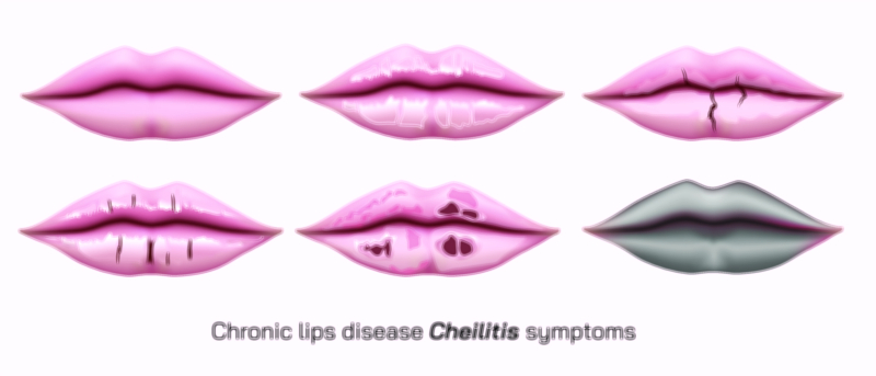
Light Therapy to Skin Vector Illustration Showing LED Treatment Benefits for Acne, Collagen, and Rejuvenation
Light therapy, commonly known as LED (Light Emitting Diode) therapy, is a non-invasive dermatological treatment that uses specific wavelengths of light to target various skin concerns. A vector illustration of light therapy applied to the skin typically highlights LED device placement, light penetration, and therapeutic effects, including acne reduction, collagen stimulation, and skin rejuvenation. By combining anatomical depiction of skin layers with visual representation of light energy, these illustrations provide a clear and educational overview of the technology and its benefits, making complex dermatological concepts accessible to medical professionals, aestheticians, and patients alike.
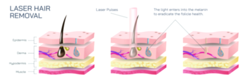
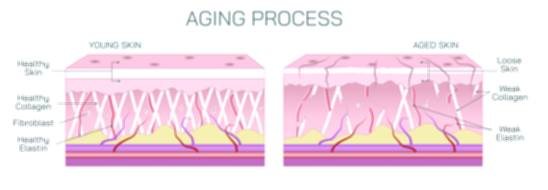
At the center of the illustration is a cross-sectional view of the skin, showing the three main layers: epidermis, dermis, and subcutaneous tissue. The epidermis is the outermost layer, protecting against environmental damage and housing the basal layer responsible for cell regeneration. The dermis contains collagen and elastin fibers, blood vessels, and hair follicles, which are key targets for rejuvenation and structural improvement. The subcutaneous layer consists primarily of fat and connective tissue, providing cushioning and thermal insulation. In the vector illustration, these layers are color-coded and labeled to provide spatial context for light penetration and its biological effects.
A central feature of LED light therapy illustrations is the depiction of different wavelengths of light and their respective skin targets. Blue light (approximately 415 nm) is shown penetrating the epidermis to target Propionibacterium acnes, the bacteria responsible for acne inflammation. Arrows or gradient beams illustrate how blue light energy disrupts bacterial activity, reducing acne lesions. Red light (around 630–660 nm) penetrates deeper into the dermis, stimulating fibroblast activity and promoting collagen and elastin production, which improves skin elasticity and reduces fine lines. Some illustrations may also include near-infrared light, which penetrates even deeper to target subcutaneous tissue, aiding in tissue repair, anti-inflammatory effects, and enhanced skin tone. Color-coded beams and arrows enhance clarity, showing wavelength-specific penetration and effects within the layered skin structure.
The vector diagram may highlight collagen stimulation in the dermis with magnified sections showing fibroblast activation, new collagen fiber formation, and tissue rejuvenation. Arrows may indicate the production and alignment of collagen, visually connecting light penetration to biological response. Similarly, acne-targeted effects in the epidermis can be illustrated with the reduction of bacteria, decreased inflammation, and normalization of sebaceous gland activity. Labels indicate each therapeutic outcome, demonstrating the targeted benefits of LED light therapy.
Additional elements often included in the illustration are LED device placement and patient positioning. A panel may show a hand-held device or a panel emitting light over facial skin, with arrows indicating the direction of energy emission. This provides a real-world context, showing how light therapy is applied clinically or cosmetically. Magnified beam diagrams may show the intensity and focus of light, emphasizing precision in targeting specific skin layers.
Vector illustrations may also incorporate comparative effects before and after treatment, highlighting improvements in acne lesions, skin texture, and firmness. Shading, color gradients, or side-by-side panels can visually communicate the benefits, such as smoother skin, reduced redness, and increased collagen density. Some diagrams may include microscopic cellular representations to show the mechanistic effects of light therapy at the cellular level, enhancing scientific understanding.
For educational purposes, arrows and labels in the illustration often indicate biological mechanisms activated by LED light, including increased fibroblast proliferation, collagen synthesis, enhanced blood circulation, reduction of inflammatory mediators, and antimicrobial effects. Visual cues such as heatmaps or glow effects can illustrate energy absorption and distribution across skin layers. This combination of anatomy and functional visualization allows viewers to connect the external application of light with internal cellular responses.
By integrating skin layers, light wavelengths, LED device placement, acne reduction, collagen stimulation, and rejuvenation effects, a vector illustration of light therapy provides a comprehensive and intuitive understanding of dermatological LED treatments. It highlights the precision of treatment, the biological mechanisms at work, and the aesthetic and therapeutic outcomes, making complex concepts visually accessible.
Ultimately, a vector illustration of skin light therapy demonstrates the interplay between technology and human physiology, showing how specific wavelengths of light target bacterial pathogens, stimulate collagen, and promote skin rejuvenation. Through labeled skin layers, color-coded light beams, directional arrows, and magnified cellular effects, the diagram transforms abstract dermatological principles into a visually engaging and educational tool, enhancing comprehension for students, clinicians, and patients interested in LED skin therapies.